. 2014 Aug 21;55(9):5979-88.
doi: 10.1167/iovs.14-15025.

Systemic administration of the antioxidant/iron chelator α-lipoic acid protects against light-induced photoreceptor degeneration in the mouse retina

Affiliations

Systemic administration of the antioxidant/iron chelator α-lipoic acid protects against light-induced photoreceptor degeneration in the mouse retina

Liangliang Zhao et al. Invest Ophthalmol Vis Sci. .

Abstract

Purpose: Oxidative stress and inflammation have key roles in the light damage (LD) model of retinal degeneration as well as in age-related macular degeneration (AMD). We sought to determine if lipoic acid (LA), an antioxidant and iron chelator, protects the retina against LD.

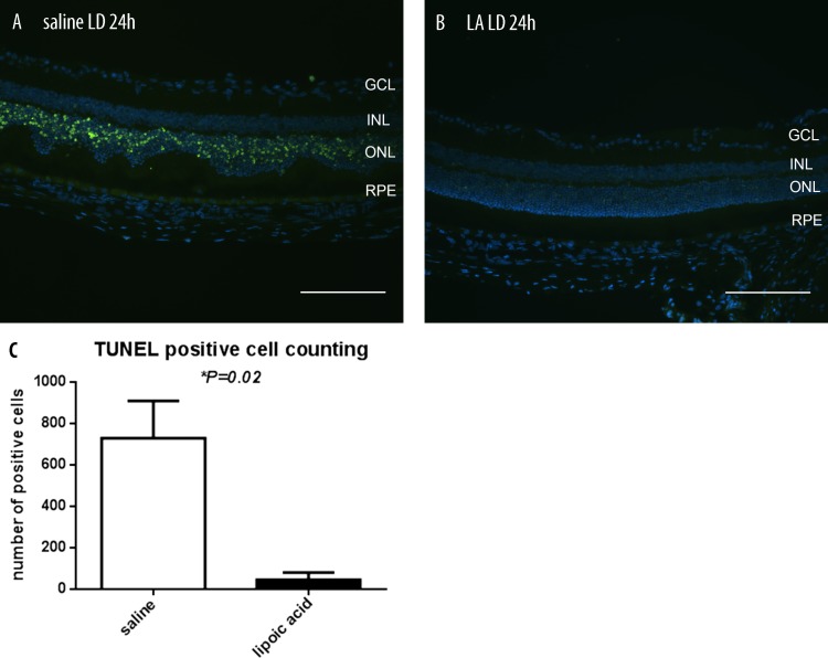
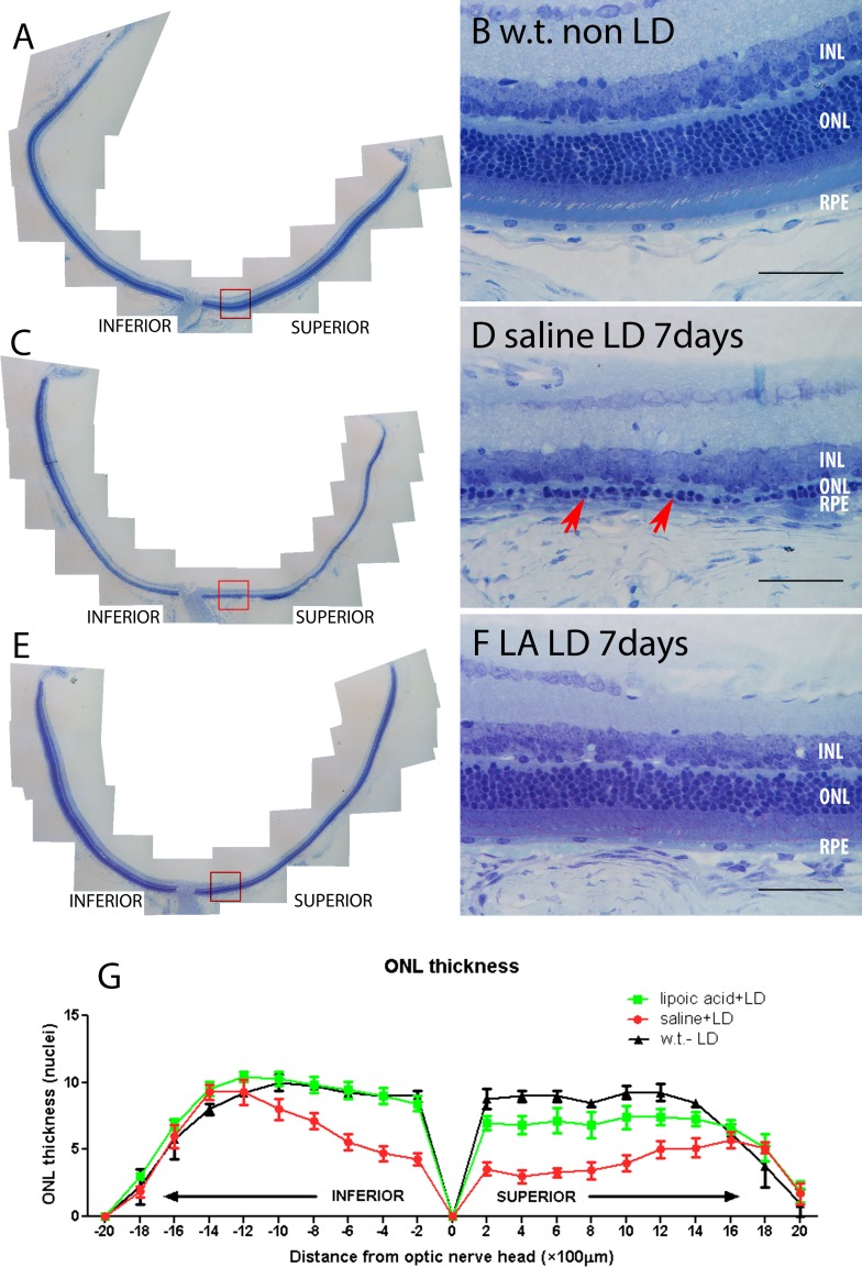
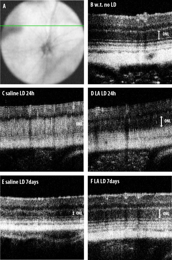
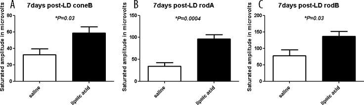
Methods: Balb/c mice were treated with LA or control saline via intraperitoneal injection, and then were placed in constant cool white light-emitting diode (LED) light (10,000 lux) for 4 hours. Retinas were evaluated at several time points after LD. Photoreceptor apoptosis was assessed using the TUNEL assay. Retinal function was analyzed via electroretinography (ERG). Retinal degeneration was assessed after LD by optical coherence tomography (OCT), TUNEL analysis, and histology. The mRNAs of several oxidative stress, inflammation, and iron-related genes were quantified by quantitative PCR (qPCR).

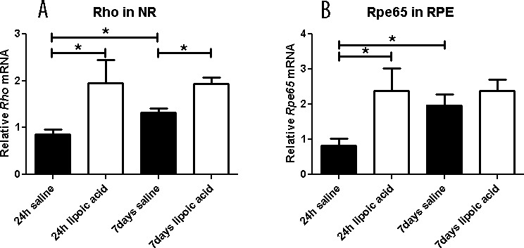
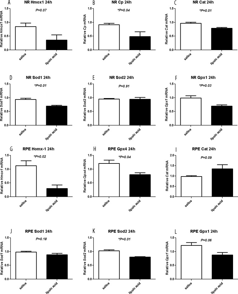
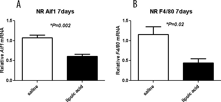
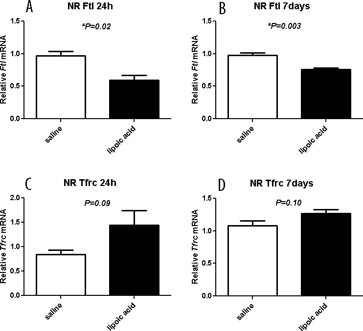
Results: The LD resulted in substantial photoreceptor-specific cell death. Dosing with LA protected photoreceptors, decreasing the numbers of TUNEL-positive photoreceptors and increasing the number of surviving photoreceptors. The retinal mRNA levels of genes indicating oxidative stress, inflammation, and iron accumulation were lower following LD in mice treated with LA than in control mice. The ERG analysis demonstrated functional protection by LA.

Conclusions: Systemic LA is protective against light-induced retinal degeneration. Since this agent already has proven protective in other retinal degeneration models, and is safe and protective against diabetic neuropathy in patients, it is worthy of consideration for a human clinical trial against retinal degeneration or AMD.

Keywords: light damage; lipoic acid; oxidative stress; retinal degeneration.


Figures

Figure 1
Figure 1
Fluorescence photomicrographs showing TUNEL label in mouse retinas. There are more TUNEL-positive photoreceptor nuclei (green) 24 hours following LD in mice treated with saline (A) compared with mice treated with LA (B). Histogram comparing numbers of TUNEL-positive photoreceptors from saline + LD (n = 3) and LA + LD (n = 3) mice. The histogram displays the mean (±SEM) of total numbers of TUNEL-positive photoreceptors counted in whole sections (C). *Significant difference (P < 0.05). GCL, ganglion cell layer; INL, inner nuclear layer. Scale bars in (A) and (B) represents 100 μm.
Figure 2
Figure 2
Photomicrographs of plastic sections of mouse retinas and plots showing morphologic protection 7 days following LD in LA + LD retinas. Retinas from mice receiving no LD (A, B), saline + LD (C, D), LA + LD (E, F). (B, D, F) Higher magnification views equidistant from the optic nerve and indicated by the red box in (A), (C), and (E), respectively. Red arrows in (D) show the thinning ONL. Plot of the thickness of the ONL 7 days after LD, measured in numbers of photoreceptor nuclei per column (G). Measurements are made in triplicate every 200 μm from the ONH. No LD (n = 4, black), saline + LD (n = 7, red), LA + LD (n = 7, green). Numbers represent mean values (±SEM). Scale bars: 100 μm (B, D, F).
Figure 3
Figure 3
The OCT images of mouse retinas. (A) Indicates the position of the line scan superior to the optic nerve. Compared to mice receiving no light damage (B), mice receiving IP saline showed increased reflectivity in the ONL 1 day after LD (C). Mice receiving IP LA had a more normal-appearing, low reflectivity ONL 1 day after LD (D). The mice receiving saline showed severe ONL thinning 7 days after LD (E), whereas mice receiving LA did not (F).
Figure 4
Figure 4
Full-field ERG responses of control saline– or LA-treated mice 7 days after light exposure. Maximum amplitude ERG responses are reported. The LA-treated mice had significantly higher amplitudes for all three wave types compared to control saline treated-animals. Numbers represent mean values (±SEM).
Figure 5
Figure 5
Graphs showing relative mRNA levels measured by qPCR. The LA treatment results in higher Rho mRNA level in NR at 1 and 7 days after LD (A) and RPE65 mRNA levels in RPE at 1 day after LD (B). In the saline group Rho mRNA levels in NR show recovery comparing 24 hours and 7 days. Similarly, in the saline group RPE65 mRNA levels in RPE show recovery comparing 24 hours and 7 days. Numbers represent mean values (±SEM).
Figure 6
Figure 6
Graphs showing relative mRNA levels measured by qPCR. The LA treatment of LD mice results in lower mRNA levels, compared to saline treatment mice, of oxidative stress markers Cp, Cat, Sod1, and Gpx1 in neural retina (B, C, D, F) and Hmox-1, Gpx4, and Sod2 in RPE (G, H, K). Saline (n = 4) and LA (n = 3) retinas are displayed as mean values (±SEM). *Significant difference (P < 0.05).
Figure 7
Figure 7
Graphs showing relative mRNA levels measured by qPCR. The LA decreases inflammation markers. Levels of Aif1 and F4/80 mRNA in neural retina are significantly diminished by the LA treatment 7 days after light exposure. Neural retina mRNA levels for the indicated genes in saline- (n = 4) and LA- (n = 4) treated mice are displayed as mean values (±SEM). *Significant difference (P < 0.05).
Figure 8
Figure 8
Graphs showing relative mRNA levels of iron handling genes measured by qPCR. The LA results in lower L-ferritin mRNA levels in neural retina at 24 hours (A) and 7 days (B) after LD. The Tfrc mRNA levels in neural retina of LA-treated mice at 24 hours (C) and 7 days (D) are not significantly different in saline- versus LA-treated mice. Saline (n = 4) and LA (n = 3) retinas are displayed as mean values (±SEM). *Significant difference (P < 0.05).

References

    1. Zarbin MA. Current concepts in the pathogenesis of age-related macular degeneration. Arch Ophthalmol. 2004; 122: 598–614 - PubMed
    1. Noell WK, Walker VS, Kang BS, Berman S. Retinal damage by light in rats. Invest Ophthalmol. 1966; 5: 450–473 - PubMed
    1. Shahinfar S, Edward DP, Tso MO. A pathologic study of photoreceptor cell death in retinal photic injury. Curr Eye Res. 1991; 10: 47–59 - PubMed
    1. Wong RW, Richa DC, Hahn P, Green WR, Dunaief JL. Iron toxicity as a potential factor in AMD. Retina Phila Pa. 2007; 27: 997–1003 - PubMed
    1. He X, Hahn P, Iacovelli J, et al. Iron homeostasis and toxicity in retinal degeneration. Prog Retin Eye Res. 2007; 26: 649–673 - PMC - PubMed

Publication types

MeSH terms