

Structural imaging measures of brain aging
- PMID: 25146995
- PMCID: PMC4163469
- DOI: 10.1007/s11065-014-9268-3
Structural imaging measures of brain aging
Abstract
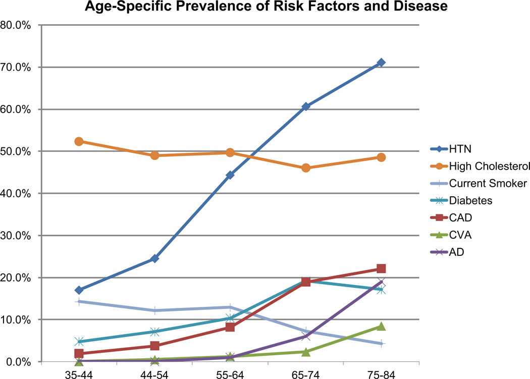
During the course of normal aging, biological changes occur in the brain that are associated with changes in cognitive ability. This review presents data from neuroimaging studies of primarily "normal" or healthy brain aging. As such, we focus on research in unimpaired or nondemented older adults, but also include findings from lifespan studies that include younger and middle aged individuals as well as from populations with prodromal or clinically symptomatic disease such as cerebrovascular or Alzheimer's disease. This review predominantly addresses structural MRI biomarkers, such as volumetric or thickness measures from anatomical images, and measures of white matter injury and integrity respectively from FLAIR or DTI, and includes complementary data from PET and cognitive or clinical testing as appropriate. The findings reveal highly consistent age-related differences in brain structure, particularly frontal lobe and medial temporal regions that are also accompanied by age-related differences in frontal and medial temporal lobe mediated cognitive abilities. Newer findings also suggest that degeneration of specific white matter tracts such as those passing through the genu and splenium of the corpus callosum may also be related to age-related differences in cognitive performance. Interpretation of these findings, however, must be tempered by the fact that comorbid diseases such as cerebrovascular and Alzheimer's disease also increase in prevalence with advancing age. As such, this review discusses challenges related to interpretation of current theories of cognitive aging in light of the common occurrence of these later-life diseases. Understanding the differences between "Normal" and "Healthy" brain aging and identifying potential modifiable risk factors for brain aging is critical to inform potential treatments to stall or reverse the effects of brain aging and possibly extend cognitive health for our aging society.
Figures
References
-
- Assaf Y, Pasternak O. Diffusion tensor imaging (DTI)-based white matter mapping in brain research: a review. Journal of Molecular Neuroscience : MN. 2008;34(1):51–61. - PubMed
-
- Boon A, Lodder J, Heuts-van Raak L, Kessels F. Silent brain infarcts in 755 consecutive patients with a first-ever supratentorial ischemic stroke. Relationship with index-stroke subtype, vascular risk factors, and mortality. Stroke. 1994;25(12):2384–2390. - PubMed
Publication types
MeSH terms
Substances
Grants and funding
LinkOut - more resources
Full Text Sources
Other Literature Sources
Medical
