. 2014 Oct;20(10):1579-92.
doi: 10.1261/rna.044933.114. Epub 2014 Aug 21.

The interaction of cytoplasmic poly(A)-binding protein with eukaryotic initiation factor 4G suppresses nonsense-mediated mRNA decay

Affiliations

The interaction of cytoplasmic poly(A)-binding protein with eukaryotic initiation factor 4G suppresses nonsense-mediated mRNA decay

Tobias Fatscher et al. RNA. 2014 Oct.

Abstract

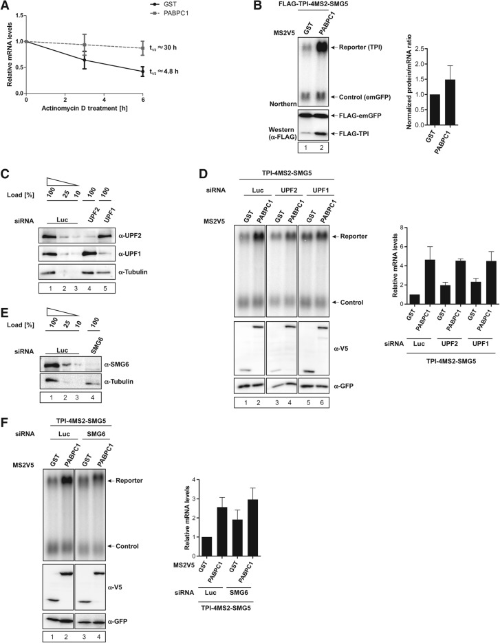
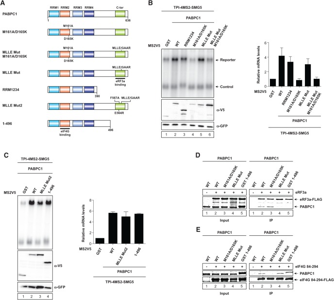
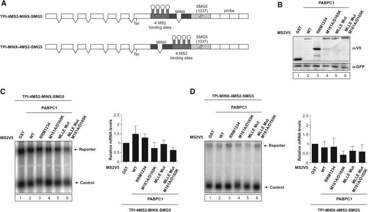
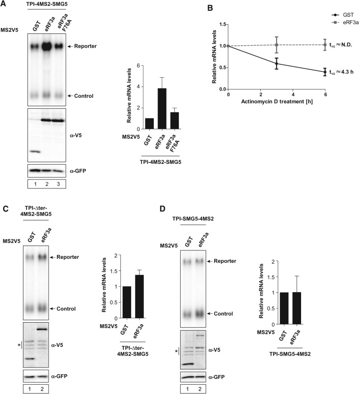
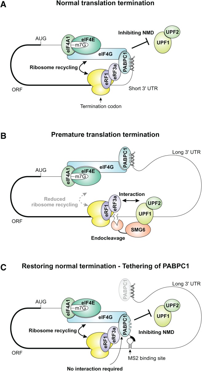
Nonsense-mediated mRNA decay (NMD) eliminates different classes of mRNA substrates including transcripts with long 3' UTRs. Current models of NMD suggest that the long physical distance between the poly(A) tail and the termination codon reduces the interaction between cytoplasmic poly(A)-binding protein (PABPC1) and the eukaryotic release factor 3a (eRF3a) during translation termination. In the absence of PABPC1 binding, eRF3a recruits the NMD factor UPF1 to the terminating ribosome, triggering mRNA degradation. Here, we have used the MS2 tethering system to investigate the suppression of NMD by PABPC1. We show that tethering of PABPC1 between the termination codon and a long 3' UTR specifically inhibits NMD-mediated mRNA degradation. Contrary to the current model, tethered PABPC1 mutants unable to interact with eRF3a still efficiently suppress NMD. We find that the interaction of PABPC1 with eukaryotic initiation factor 4G (eIF4G), which mediates the circularization of mRNAs, is essential for NMD inhibition by tethered PABPC1. Furthermore, recruiting either eRF3a or eIF4G in proximity to an upstream termination codon antagonizes NMD. While tethering of an eRF3a mutant unable to interact with PABPC1 fails to suppress NMD, tethered eIF4G inhibits NMD in a PABPC1-independent manner, indicating a sequential arrangement of NMD antagonizing factors. In conclusion, our results establish a previously unrecognized link between translation termination, mRNA circularization, and NMD suppression, thereby suggesting a revised model for the activation of NMD at termination codons upstream of long 3' UTR.

Keywords: NMD; PABPC1; eIF4G; ribosome recycling; translation termination.


Figures

FIGURE 1.
FIGURE 1.
Tethering PABPC1 to a reporter mRNA containing a long 3′ UTR increases mRNA abundance. (A) Schematic representation of the triosephosphate isomerase (TPI) reporter constructs. White boxes depict exons, introns are shown as two connecting black lines, and Northern probe binding sites as white boxes without intron lines. Gray boxes represent MS2-binding area with MS2-stem–loops shown in black. The SMG5 3′ UTR is depicted as a light-gray box and the length in nucleotides is shown in brackets. (BF) Northern blot analysis of total RNA extracted from HeLa cells transfected with plasmids expressing the indicated TPI reporter mRNA and MS2V5- or FLAG-tagged fusion proteins. A β-globin construct was cotransfected as control. Protein expression was detected by immunoblotting with α-V5 or α-FLAG antibody. Cotransfected GFP served as a loading control. Asterisks indicate unspecific bands (C,D). mRNA levels were normalized to MS2V5-GST (BD,F) or pCI-FLAG (E). Bars represent the mean values of mRNA levels±SD upon tethering MS2V5-GST or MS2V5-PABPC1 (BD,F), or pCI-FLAG and FLAG-PABPC1 (E). Concentrations of MS2V5-tagged protein expressing plasmids were increased from 1 µg (F, lanes 1,3,5,7) to 3 µg (F, lanes 2,4,6,8).
FIGURE 2.
FIGURE 2.
PABPC1 stabilizes reporter mRNA by suppressing NMD. (A) HeLa cells expressing reporter (TPI-4MS2-SMG5) and control mRNA, as well as MS2V5-tagged GST or PABPC1, were treated with Actinomycin D (5 µg/mL final concentration) for the indicated time prior to harvesting. Reporter mRNA levels were quantified by Northern blotting, normalized to control mRNA and GST control tethering, and plotted against time of Actinomycin D treatment. (B) Tethering of MS2V5-GST and -PABPC1 in HeLa cells cotransfected with N-terminally FLAG-tagged TPI-4MS2-SMG5 reporter and FLAG-tagged emGFP control expressing vectors. Both reporter and control mRNA contained heterologous binding sites in the 3′ UTR that enable the detection with the same Northern probe. Northern blot (top) and α-FLAG Western blot (bottom) analyses are shown. The signals for emGFP and TPI in Northern and Western blot experiments were quantified, normalized to the GST control lane, and the final ratio was calculated by normalizing protein expression levels to the respective mRNA expression levels. (C,E) siRNA-mediated knockdown of UPF2, UPF1, and SMG6. HeLa cells were transfected with siRNAs targeting UPF2, UPF1, SMG6, or Luciferase (negative control). The knockdown efficiency was assessed by immunoblotting with UPF2, UPF1, and SMG6-specific antibodies. Tubulin served as a loading control. (D,F) Northern blot analysis of TPI-4MS2-SMG5 reporter mRNA with β-globin mRNA as control in UPF2 (D, lanes 3,4), UPF1 (D, lanes 5,6), and SMG6 (F, lanes 3,4) knockdown and control (D,F, lanes 1,2) cells. HeLa cells were transfected with plasmids expressing MS2V5-GST and -PABPC1 proteins. Protein expression was detected by immunoblotting with a V5 antibody. GFP served as a loading control. mRNA levels were normalized to MS2V5-GST. Bars represent the mean values of mRNA levels ±SD upon tethering MS2V5-GST and -PABPC1 fusion proteins.
FIGURE 3.
FIGURE 3.
PABPC1 interaction with eIF4G, but not eRF3a, is essential for NMD suppression. (A) Schematic representation of PABPC1 domains and mutants. RNA recognition motifs (RRMs) and C-terminal region (C-ter) of PABPC1 are highlighted. Point mutations and binding sites are indicated. (B,C) HeLa cells were transfected with plasmids expressing the indicated MS2V5-tagged fusion proteins and the indicated TPI reporter mRNA. Northern blot analysis was performed and cotransfected β-globin mRNA construct served as control. Protein expression was detected by immunoblotting with a V5 antibody. GFP served as a loading control. mRNA levels were normalized to MS2V5-GST. Bars represent mean values of mRNA levels ±SD upon tethering of different MS2V5-tagged fusion proteins. (D,E) Pull-down assays of in vitro interaction studies using PABPC1 mutants and FLAG-tagged eRF3a or eIF4G 84-294. Proteins were visualized with Coomassie Brilliant Blue.
FIGURE 4.
FIGURE 4.
PABPC1 primarily functions as a suppressor in EJC-independent NMD. (A) Schematic representation of the TPI reporters as in Figure 1A. Dark-gray boxes indicate intron-containing MINX cassette. (B) Representative Western blot showing MS2V5-tagged protein expression, as analyzed by immunoblotting with a V5 antibody. (C,D) HeLa cells were transfected with plasmids expressing the indicated MS2V5-tagged fusion proteins and the indicated TPI reporter mRNA. Northern blot analysis was performed and cotransfected β-globin mRNA construct served as control. mRNA levels were normalized to MS2V5-GST. Bars represent mean values of mRNA levels ±SD upon tethering of different MS2V5-tagged fusion proteins.
FIGURE 5.
FIGURE 5.
Tethered eRF3a unable to interact with PABPC1 fails to stabilize reporter mRNA levels. (A,C,D) HeLa cells were transfected with plasmids expressing the indicated MS2V5-tagged fusion proteins and the indicated TPI reporter mRNA. Northern blot analysis was performed and cotransfected β-globin mRNA construct served as control. Protein expression was detected by immunoblotting with a V5 antibody. GFP served as a loading control. Asterisks indicate unspecific bands (C,D). mRNA levels were normalized to MS2V5-GST. Bars represent mean values of mRNA levels ±SD upon tethering of different MS2V5-tagged fusion proteins. (B) HeLa cells expressing MS2V5-tagged GST or eRF3a were treated with Actinomycin D for the indicated time. mRNA levels were quantified after Northern blot analysis and plotted as described in Figure 2.
FIGURE 6.
FIGURE 6.
The interaction with PABPC1 is dispensable for NMD suppression by tethered eIF4G. (A) Illustration of eIF4G domain architecture and mutants. MIF4G, MI, and W2 domains are highlighted. Point mutations and binding sites are indicated. (B,E,F) HeLa cells were transfected with plasmids expressing the indicated MS2V5-tagged fusion proteins and the indicated TPI reporter mRNA. Northern blot analysis was performed and cotransfected β-globin mRNA construct served as control. Protein expression was detected by immunoblotting with a V5 antibody. GFP served as a loading control. Asterisk indicates an unspecific band (B). mRNA levels were normalized to MS2V5-GST. Bars represent mean values of mRNA levels ±SD upon tethering of different MS2V5-tagged fusion proteins. (C) Pull-down assays of in vitro interaction studies using PABPC1 and FLAG-tagged eIF4G 84-294 WT or KRERK mutant. Proteins were visualized with Coomassie Brilliant Blue. (D) HeLa cells expressing MS2V5-tagged GST or eIF4G ΔN83 were treated with Actinomycin D for the indicated time. mRNA levels were quantified after Northern blot analysis and plotted as described in Figure 2. (G) Representative Western blot showing MS2V5-tagged protein expression, as analyzed by immunoblotting with a V5 antibody. GFP served as a loading control. Asterisk indicates an unspecific band.
FIGURE 7.
FIGURE 7.
Model for a link between eIF4G-mediated ribosome recycling and NMD inhibition. (A) Normal translation termination and ribosome recycling of a short 3′ UTR-containing mRNA is enabled by PABPC1 interacting with both eRF3a and eIF4G, thereby preventing NMD. (B) Aberrant translation termination of long 3′ UTR-containing mRNAs activates NMD. The interaction of PABPC1 with eRF3a is decreased due to a large physical distance, preventing efficient ribosome recycling by eIF4G. Consequently, UPF1 is postulated to interact with eRF3a and elicit NMD. (C) Tethered PABPC1 inhibits NMD of a long 3′ UTR-containing substrate by bringing eIF4G in close proximity to the terminating ribosome. This proximity promotes a proper translation termination event and facilitates ribosome recycling, thereby antagonizing NMD activation.

References

    1. Amrani N, Ganesan R, Kervestin S, Mangus DA, Ghosh S, Jacobson A 2004. A faux 3′-UTR promotes aberrant termination and triggers nonsense-mediated mRNA decay. Nature 432: 112–118 - PubMed
    1. Amrani N, Ghosh S, Mangus DA, Jacobson A 2008. Translation factors promote the formation of two states of the closed-loop mRNP. Nature 453: 1276–1280 - PMC - PubMed
    1. Behm-Ansmant I, Gatfield D, Rehwinkel J, Hilgers V, Izaurralde E 2007. A conserved role for cytoplasmic poly(A)-binding protein 1 (PABPC1) in nonsense-mediated mRNA decay. EMBO J 26: 1591–1601 - PMC - PubMed
    1. Bhuvanagiri M, Schlitter AM, Hentze MW, Kulozik AE 2010. NMD: RNA biology meets human genetic medicine. Biochem J 430: 365–377 - PubMed
    1. Burgess HM, Richardson WA, Anderson RC, Salaun C, Graham SV, Gray NK 2011. Nuclear relocalisation of cytoplasmic poly(A)-binding proteins PABP1 and PABP4 in response to UV irradiation reveals mRNA-dependent export of metazoan PABPs. J Cell Sci 124: 3344–3355 - PMC - PubMed

Publication types

MeSH terms