

Review
doi: 10.3402/dfa.v5.24445.
eCollection 2014.
Osteomyelitis in the diabetic foot
Affiliations
- PMID: 25147627
- PMCID: PMC4119293
- DOI: 10.3402/dfa.v5.24445
Item in Clipboard
Review
Osteomyelitis in the diabetic foot
Diabet Foot Ankle.
.
Abstract
Osteomyelitis (OM) is a common complication of diabetic foot ulcers and/or diabetic foot infections. This review article discusses the clinical presentation, diagnosis, and treatment of OM in the diabetic foot. Clinical features that point to the possibility of OM include the presence of exposed bone in the depth of a diabetic foot ulcer. Medical imaging studies include plain radiographs, magnetic resonance imaging, and bone scintigraphy. A high index of suspicion is also required to make the diagnosis of OM in the diabetic foot combined with clinical and radiological studies.
Keywords: amputation; antibiotics; diabetic foot; infection; osteomyelitis; ulcer.
Figures
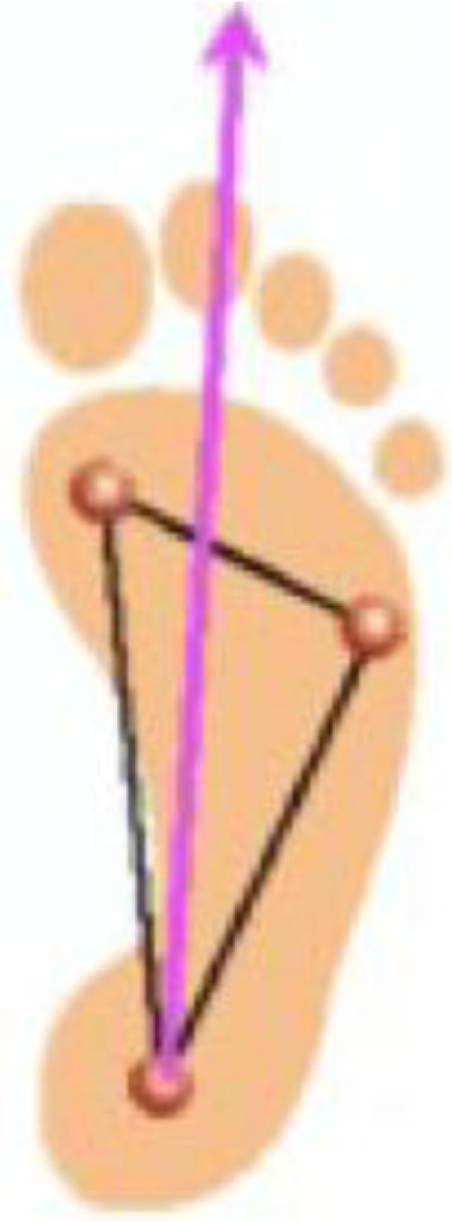
Diagram of weight-bearing tripod of the foot.
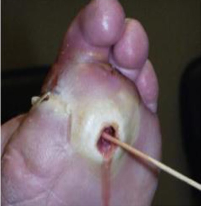
Probe-to-bone test for osteomyelitis in the diabetic foot.
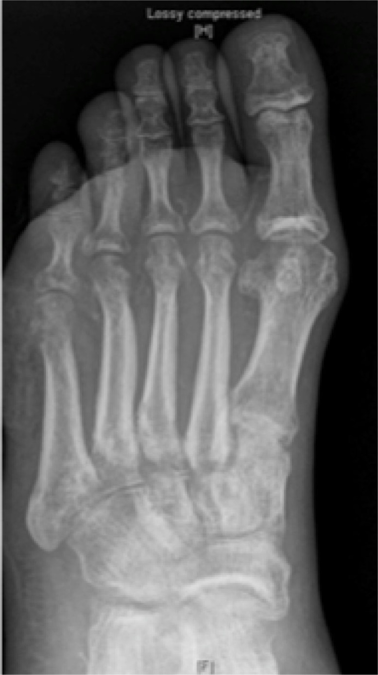
Plain radiograph. Evidence of cortical erosion of the fifth metatarsal head in a patient with osteomyelitis.
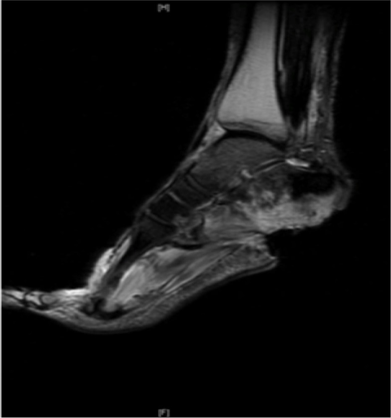
MRI. T2-weighted image – irregular ‘whitening’ of calcaneum suggests edema and osteomyelitis in a patient with a calcaneal ulcer.

Bone scan. Bone scan showing increased uptake localized to the base of the fifth metatarsal, indicating osteomyelitis.

Showing bones removed during a second ray amputation (second toe and partial resection of the second metatarsal).
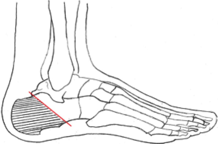
Diagram showing the bone resection for partial calcanectomy.
References
-
- Mutluoglu M, Sivrioglu AK, Eroglu M, Uzun G, Turhan V, Ay H, et al. The implications of the presence of osteomyelitis on outcomes of infected diabetic foot wounds. Scand J Infect Dis. 2013;45:497–503. - PubMed
-
- Game FL. Osteomyelitis in the diabetic foot: diagnosis and management. Med Clin North Am. 2013;97:947–56. - PubMed
-
- Ramsey SD, Newton K, Blough D, McCulloch DK, Sandhu N, Reiber GE, et al. Incidence, outcomes, and cost of foot ulcers in patients with diabetes. Diabetes Care. 1999;22:382–7. - PubMed
-
- Lavery LA, Peters EJ, Armstrong DG, Wendel CS, Murdoch DP, Lipsky BA. Risk factors for developing osteomyelitis in patients with diabetic foot wounds. Diabetes Res Clin Pract. 2009;83:347–52. - PubMed
-
- Newman LG, Waller J, Palestro CJ, Schwartz M, Klein MJ, Hermann G, et al. Unsuspected osteomyelitis in diabetic foot ulcers. Diagnosis and monitoring by leukocyte scanning with indium in 111 oxyquinoline. JAMA. 1991;266:1246–51. - PubMed
Publication types
LinkOut - more resources
Full Text Sources
Other Literature Sources