Review
. 2014 Aug;12(6):315-41.
doi: 10.1089/adt.2014.587.

Assays for the identification and prioritization of drug candidates for spinal muscular atrophy

Affiliations
Review

Assays for the identification and prioritization of drug candidates for spinal muscular atrophy

Jonathan J Cherry et al. Assay Drug Dev Technol. 2014 Aug.

Abstract

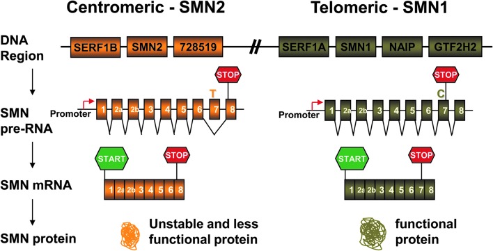
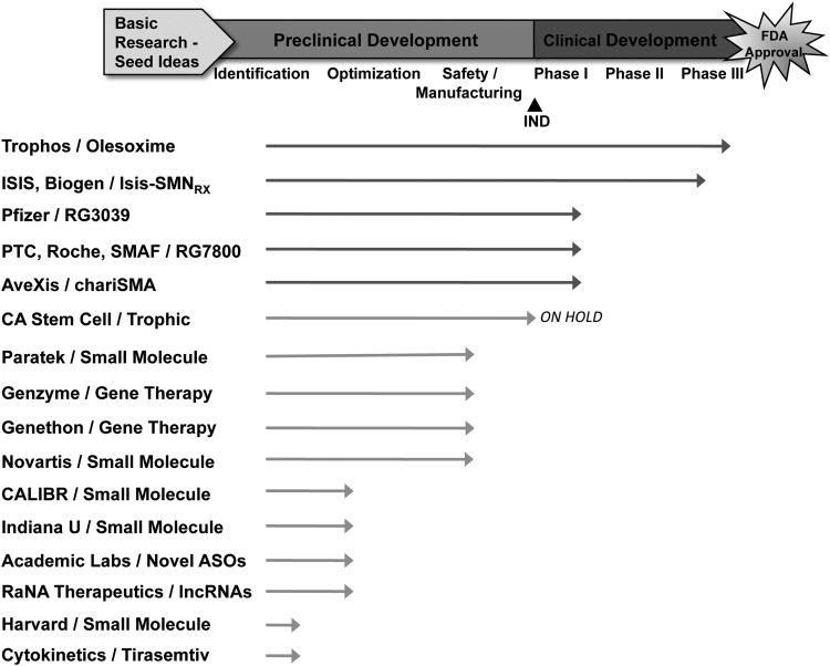
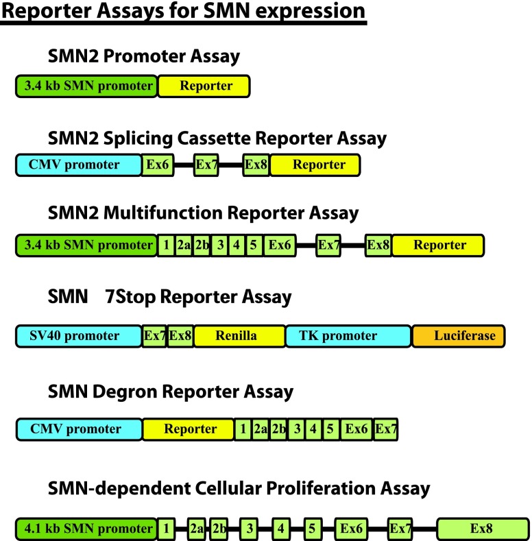
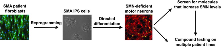
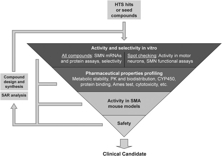
Spinal muscular atrophy (SMA) is an autosomal recessive genetic disorder resulting in degeneration of α-motor neurons of the anterior horn and proximal muscle weakness. It is the leading cause of genetic mortality in children younger than 2 years. It affects ∼1 in 11,000 live births. In 95% of cases, SMA is caused by homozygous deletion of the SMN1 gene. In addition, all patients possess at least one copy of an almost identical gene called SMN2. A single point mutation in exon 7 of the SMN2 gene results in the production of low levels of full-length survival of motor neuron (SMN) protein at amounts insufficient to compensate for the loss of the SMN1 gene. Although no drug treatments are available for SMA, a number of drug discovery and development programs are ongoing, with several currently in clinical trials. This review describes the assays used to identify candidate drugs for SMA that modulate SMN2 gene expression by various means. Specifically, it discusses the use of high-throughput screening to identify candidate molecules from primary screens, as well as the technical aspects of a number of widely used secondary assays to assess SMN messenger ribonucleic acid (mRNA) and protein expression, localization, and function. Finally, it describes the process of iterative drug optimization utilized during preclinical SMA drug development to identify clinical candidates for testing in human clinical trials.


Figures

<b>Fig. 1.</b>
Fig. 1.
Splicing of SMN1 and SMN2. The genomic regions of the SMN1 and SMN2 genes are drawn, as shown at www.ncbi.nlm.nih.gov/gene/. The major difference between the two SMN gene copies is the C (SMN1) or T (SMN2) nucleotide change at position 6 in exon 7 of the two genes. This single-nucleotide change prevents the binding of the SR protein and splicing activator ASF/SF2, in addition to creating an inhibitory binding element for proteins such as hnRNPA1 and Sam68. Because of this, SMN2 primarily produces messenger ribonucleic acid (mRNA) that excludes exon 7 and results in truncated and unstable SMN protein. However, SMN1 mostly produces mRNA that includes exon 7 and results in stable full-length SMN protein. Adapted with permission from the Families of SMA publication “The Genetics of Spinal Muscular Atrophy.” ASF/SF2, alternative splicing factor 1/pre-mRNA-splicing factor 2; SMN, survival of motor neuron.
<b>Fig. 2.</b>
Fig. 2.
SMA drug pipeline. The status of known candidate therapies being assessed for the treatment of SMA. Status of the compounds is estimated. The academic laboratories working on novel ASOs include those of Dr. Ravindra Singh at Iowa State University, Dr. Christian Lorson at University of Missouri, and Dr. Arthur Burghes at Ohio State University. lncRNAs, long noncoding RNAs; IND, Investigational New Drug; ASO, antisense oligonucleotide; SMAF, Spinal Muscular Atrophy Foundation; U, University; SMA, spinal muscular atrophy.
<b>Fig. 3.</b>
Fig. 3.
Schematic representation of SMN2 reporter constructs. SMN sequences are colored green. Non-SMN promoter elements are blue. Reporter genes (yellow and orange) vary in different versions of each construct (see Table 4 for details).
<b>Fig. 4.</b>
Fig. 4.
Schematic outline of the use of iPS cells in drug screening for SMA. SMA patient fibroblasts (green, SMN; blue, DAPI) can be reprogrammed to iPS cells (shown in brightfield). These pluripotent cells can be differentiated into motor neurons (red, neuronal marker Tuj1; green, motor neuron marker Islet1). These SMA patient-derived motor neurons can then be used for drug screening as well as for testing therapeutic candidates across multiple patient lines. iPS, induced pluripotent stem.
<b>Fig. 5.</b>
Fig. 5.
A screening cascade for the identification of SMN2 expression modulators. Testing is organized in a sequential manner, with high-throughput in vitro activity and selectivity assays residing in the top tier, followed by profiling for drug-like properties and penetration into CNS and other target tissues in the second tier, determination of activity in SMA mouse models in the third tier, and assessment of in vivo safety in the fourth tier of testing. Predefined metrics need to be satisfied for progression between individual tiers. Data are collected at every tier and analyzed to build SAR models, as indicated by the information flow arrows on the left. New structural analogs with improved properties are generated via medicinal chemistry approaches and enter the screening funnel from the top. When a compound satisfying most of the product profile criteria is identified, the selected clinical candidate can progress into human clinical trials. CNS, central nervous system; SAR, structure–activity relationship.

References

    1. Crawford TO: From enigmatic to problematic: the new molecular genetics of childhood spinal muscular atrophy. Neurology 1996;46:335–340 - PubMed
    1. McAndrew PE, Parsons DW, Simard LR, et al. : Identification of proximal spinal muscular atrophy carriers and patients by analysis of SMNT and SMNC gene copy number. Am J Hum Genet 1997;60:1411–1422 - PMC - PubMed
    1. Sugarman EA, Nagan N, Zhu H, et al. : Pan-ethnic carrier screening and prenatal diagnosis for spinal muscular atrophy: clinical laboratory analysis of >72,400 specimens. Eur J Hum Genet 2012;20:27–32 - PMC - PubMed
    1. Pearn J: Autosomal dominant spinal muscular atrophy: a clinical and genetic study. J Neurol Sci 1978;38:263–275 - PubMed
    1. Wang CH, Finkel RS, Bertini ES, et al. : Consensus statement for standard of care in spinal muscular atrophy. J Child Neurol 2007;22:1027–1049 - PubMed

MeSH terms

Substances