. 2014 Nov 21;9(11):2508-16.
doi: 10.1021/cb500236y. Epub 2014 Aug 28.

Structural basis for the inhibition of HSP70 and DnaK chaperones by small-molecule targeting of a C-terminal allosteric pocket

Affiliations

Structural basis for the inhibition of HSP70 and DnaK chaperones by small-molecule targeting of a C-terminal allosteric pocket

Julia I-Ju Leu et al. ACS Chem Biol. .

Abstract

The stress-inducible mammalian heat shock protein 70 (HSP70) and its bacterial orthologue DnaK are highly conserved nucleotide binding molecular chaperones. They represent critical regulators of cellular proteostasis, especially during conditions of enhanced stress. Cancer cells rely on HSP70 for survival, and this chaperone represents an attractive new therapeutic target. We have used a structure-activity approach and biophysical methods to characterize a class of inhibitors that bind to a unique allosteric site within the C-terminus of HSP70 and DnaK. Data from X-ray crystallography together with isothermal titration calorimetry, mutagenesis, and cell-based assays indicate that these inhibitors bind to a previously unappreciated allosteric pocket formed within the non-ATP-bound protein state. Moreover, binding of inhibitor alters the local protein conformation, resulting in reduced chaperone-client interactions and impairment of proteostasis. Our findings thereby provide a new chemical scaffold and target platform for both HSP70 and DnaK; these will be important tools with which to interrogate chaperone function and to aid ongoing efforts to optimize potency and efficacy in developing modulators of these chaperones for therapeutic use.


Figures

Figure 1
Figure 1
PET-16 is cytotoxic to human tumor cells and inhibits the growth of E. coli. (A) Chemical structures of PES, B-PES, and PET-16. The major functional groups are indicated. (B) MTT assays of human melanoma cell lines treated with the indicated concentrations of PES or PET-16 for 72 h. The corresponding cell survival is normalized to vehicle (DMSO) treatment. Average and standard deviation (s.d.) from three independent experiments are shown. (C) Human melanoma (A875) cells and nontransformed human (IMR90) fibroblasts were treated with DMSO, 10 μM PES, or 10 μM PET-16 for 24 h. The data shown are representative of four independent experiments. (D) Growth of E. coli DH5α treated with different concentrations of PES or PET-16 for 6 h at 43 °C. Error bars represent the s.d. of four independent experiments. (E) Growth of E. coli DH5α treated with DMSO, 30 μM PET-16, or 30 μM TPP for 6 h at 43 °C. Error bars represent the s.d. of four independent experiments.
Figure 2
Figure 2
PET-16 binds directly to ADP-bound HSP70 and DnaK. (A–H) Representative ITC assays of the indicated compounds (NRLLLTG, PES or PET-16) with purified HSP70-ADP, DnaK-ADP, or HSP70-ATP proteins. The data shown are representative of three independent experiments. The reported dissociation constants are averages and standard deviations from three independent experiments. (I and J) Representative ITC binding curves obtained for the interaction between the HSP70 (aa 386–616) protein and PET-16 (I) or TPP (J). (K) HSP70 protein was preincubated with ADP and PES for 1 h. The mixture was titrated into the sample cell containing PET-16. The reported dissociation constants are averages and standard deviations from three independent experiments. (L) For competition studies, human lung carcinoma (H1299) cells were pretreated with DMSO or excess (8×) PET-16 for 1 h prior to the addition of 20 μM B-PES for 5 h and examined for the expression of HSP70 or BCL-xL. B-PES-containing complexes were captured by Avidin resins and immunoblotted either with anti-HSP70 or anti-BCL-xL antibody. As shown, PET-16 inhibits the subsequent interaction of HSP70 with B-PES.
Figure 3
Figure 3
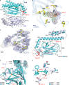
X-ray crystal structure of the DnaK-PET-16 complex. (A) Overall structure of the DnaK-PET-16 complex. The major domains and secondary structural elements are labeled. PET-16, in MolB, is shown in red. (B) Electron density map corresponding to PET-16 in the DnaK-PET-16 cocrystal structure. The 2Fo – Fc electron density map of the refined structure corresponding to PET-16 contoured at 1.0 σ is shown in blue. The Fo – Fc difference map prior to introducing PET-16 into the model is contoured at 3.0 σ and shown in green; there is no contribution from PET-16 in this map. Fo – Fc PET-16 omit map contoured at 3.0 σ is shown in red. The side chain of key PET-16 contacting residues and Gly482 are shown in stick format and labeled. The PET-16 compound is shown as a stick model in gray. Note that PET-16 binds to a pocket formed by strand β1 (L399) and loops LL,1 (L392, P396), L6,7 (G482), and Lα,β (A503 and S504). (C) Electron density map corresponding to MolA of the DnaK-PET-16 cocrystal structure. The Fo – Fc map, corresponding to the PET-16 binding site in MolB, contoured at 3.0 σ, is shown in green. The 2Fo – Fc electron density map of strand β1 (L399) and loops LL,1 (L392, P396), L6,7 (G482), and Lα,β (A503 and S504) in MolA contoured at 1.5 σ is shown in blue. Key PET-16 contacting residues are shown in yellow stick format and labeled. The electron density corresponding to PET-16, as noted in Figure 3B, was not observed in the refined structure of MolA. (D) Structural alignment of MolA with MolB. The PET-16 compound is shown in red stick format and labeled. (E and F) Structural alignment of MolA with MolB. Structural differences noted in strand β1 and loops LL,1 and Lα,β of MolA and MolB are illustrated.
Figure 4
Figure 4
Mutation analyses support the DnaK-PET-16 cocrystal structure. (A) The binding pocket for PET-16 in the DnaK-PET-16 structure. The key residues that contact PET-16 in DnaK are highlighted and labeled. (B) PET-16 and PES bind directly to the C-terminal domain of ADP-bound DnaK and HSP70. ITC-derived binding constants (Kd values) from assays of PES and PET-16 incubated with wild-type and mutated full-length ADP-DnaK (top panel) and ADP-HSP70 proteins (bottom panel). The reported dissociation constants are averages and standard deviations from three independent experiments. (C and D) Pull-down assays using H1299 cells transfected with the indicated HA-tagged HSP70 constructs followed by treatment with 20 μM B-PES for 5 h. B-PES-containing complexes were captured by avidin beads, eluted, and detected with an anti-HA antibody following immunoblotting. Input is shown on the top panel; immunoprecipitation (IP) with avidin is depicted on the bottom.
Figure 5
Figure 5
PET-16 impairs chaperone–client interaction and proteostasis. (A and B) Representative ITC-binding isotherms were recorded for the full-length recombinant human HSP70 proteins pretreated for 1 h with ADP and PET-16 (A) or with ADP and PES (B) and then titrated into a solution of substrate peptide NRLLLTG. The data shown here are representative of three independent experiments. (C and D) Whole-cell extracts (WCE) prepared from human melanoma A875 (C) or 451 Lu (D) cells treated with DMSO, PES, or PET-16 were immunoprecipitated using an anti-HSP70 antibody. The excised band of ∼70 kDa shown in the Coomassie gel is HSP70, as confirmed by liquid chromatography–tandem mass spectrometry analysis. (E) EM analysis provides evidence of altered autophagy in PET-16 treated tumor cells, with the presence of autophagosomes, accumulation of vacuoles, and the appearance of granular and aggregated masses. Note the general absence of obvious nuclei in PET-16 treated tumor cells. (F) Human melanoma 451 Lu cells were treated with DMSO, PES, or PET-16 for 24 h. Cells were harvested in 1% NP40-containing lysis buffer, fractionated into detergent-soluble and detergent-insoluble preparations, and assayed by Western blot for ubiquitin. (G) Human H1299 lung carcinoma cells, transfected with a negative shRNA (sh-Negative) or with HSP70 shRNAs (sh-HSP70), were treated with DMSO or PET-16, as indicated. Proteins were assayed by Western blot for ubiquitin in the detergent-insoluble fraction and for HSP70 and HSC70 in the detergent-soluble fraction.

References

    1. Mayer M.-P.; Bukau B. (2005) Hsp70 chaperones: cellular functions and molecular mechanism. Cell. Mol. Life Sci. 62, 670–684. - PMC - PubMed
    1. Daugaard M.; Rohde M.; Jäättelä M. (2007) The heat shock protein 70 family: highly homologous proteins with overlapping and distinct functions. FEBS Lett. 581, 3702–3710. - PubMed
    1. Kim Y.-E.; Hipp M.-S.; Hayer-Hartl A.-B.-M.; Hartl F.-U. (2013) Molecular chaperone functions in protein folding and proteostasis. Annu. Rev. Biochem. 82, 323–355. - PubMed
    1. Mayer M.-P. (2013) Hsp70 chaperone dynamics and molecular mechanism. Trends Biochem. Sci. 38, 507–514. - PubMed
    1. Murphy M.-E. (2013) The HSP70 family and cancer. Carcinogenesis 34, 1181–1188. - PMC - PubMed

Publication types

MeSH terms

Substances