. 2014 Aug 22;9(8):e104391.
doi: 10.1371/journal.pone.0104391. eCollection 2014.

Eukaryotic initiation factor 4G suppresses nonsense-mediated mRNA decay by two genetically separable mechanisms

Affiliations

Eukaryotic initiation factor 4G suppresses nonsense-mediated mRNA decay by two genetically separable mechanisms

Raphael Joncourt et al. PLoS One. .

Abstract

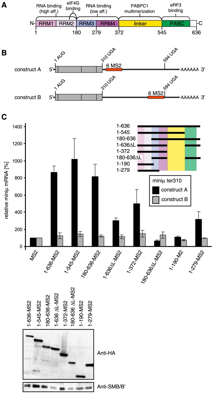
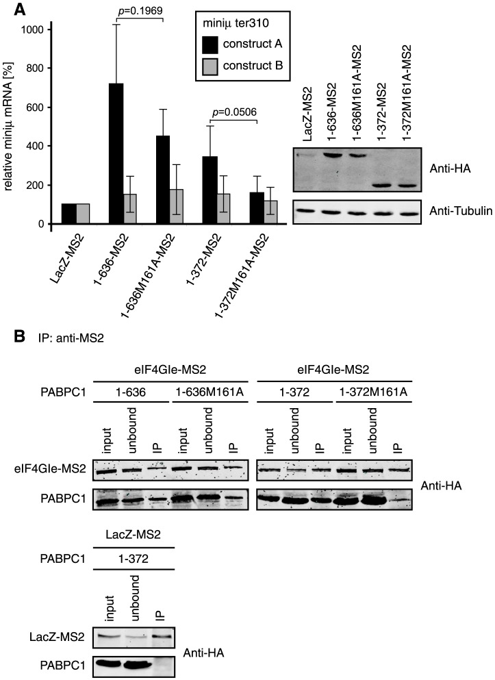
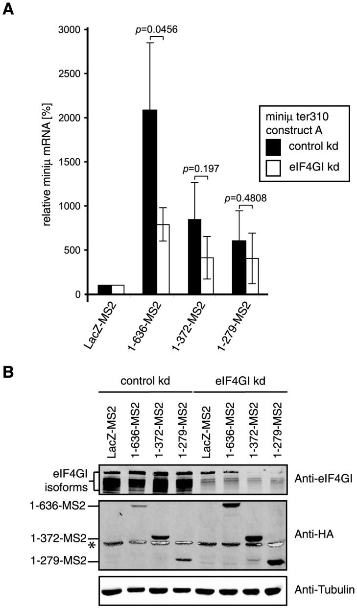
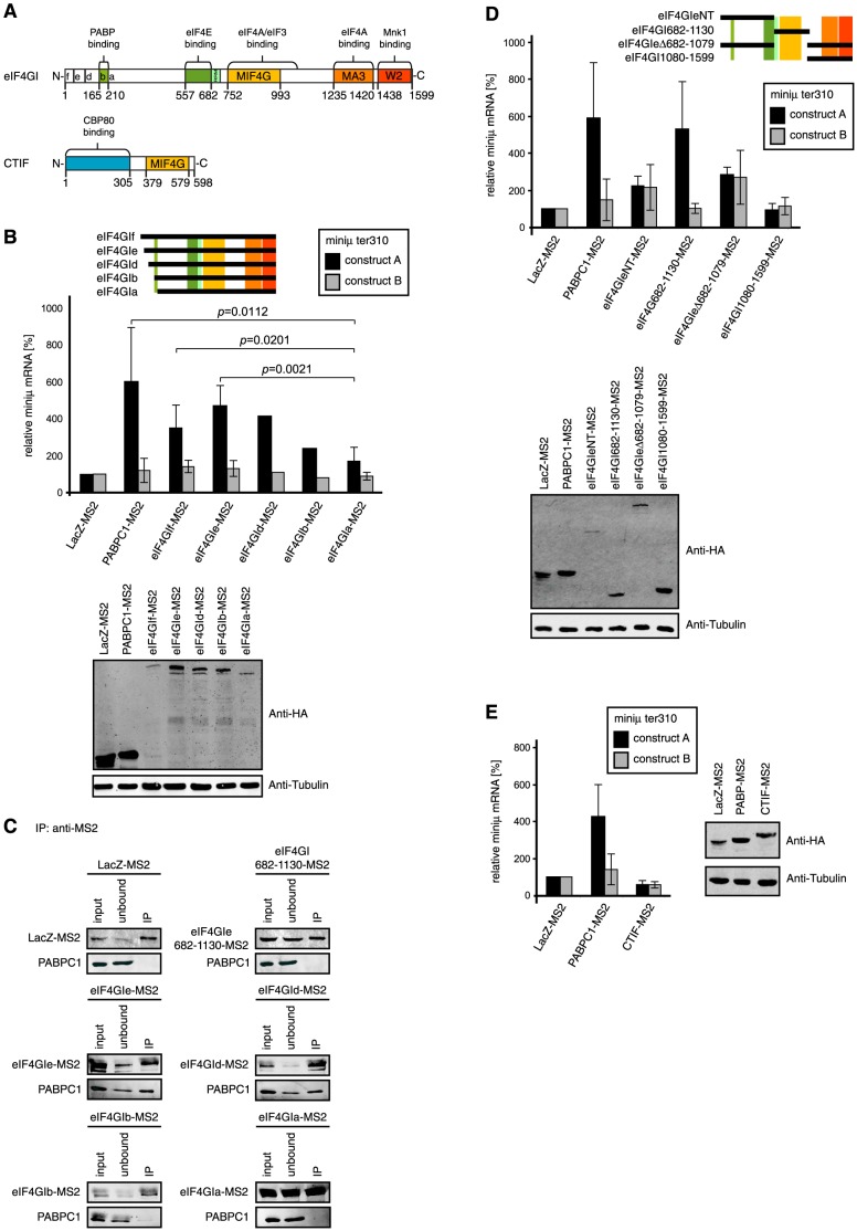
Nonsense-mediated mRNA decay (NMD), which is best known for degrading mRNAs with premature termination codons (PTCs), is thought to be triggered by aberrant translation termination at stop codons located in an environment of the mRNP that is devoid of signals necessary for proper termination. In mammals, the cytoplasmic poly(A)-binding protein 1 (PABPC1) has been reported to promote correct termination and therewith antagonize NMD by interacting with the eukaryotic release factors 1 (eRF1) and 3 (eRF3). Using tethering assays in which proteins of interest are recruited as MS2 fusions to a NMD reporter transcript, we show that the three N-terminal RNA recognition motifs (RRMs) of PABPC1 are sufficient to antagonize NMD, while the eRF3-interacting C-terminal domain is dispensable. The RRM1-3 portion of PABPC1 interacts with eukaryotic initiation factor 4G (eIF4G) and tethering of eIF4G to the NMD reporter also suppresses NMD. We identified the interactions of the eIF4G N-terminus with PABPC1 and the eIF4G core domain with eIF3 as two genetically separable features that independently enable tethered eIF4G to inhibit NMD. Collectively, our results reveal a function of PABPC1, eIF4G and eIF3 in translation termination and NMD suppression, and they provide additional evidence for a tight coupling between translation termination and initiation.


Conflict of interest statement

Competing Interests: The authors have declared that no competing interests exist.

Figures

Figure 1
Figure 1. The C-terminal domain of PABPC1 is dispensable for inhibition of NMD, RRMs1-3 are sufficient, and RRMs1-2 are necessary.
(A) Schematic representation of the eukaryotic cytoplasmic poly(A) binding protein (PABPC1). Domains are color-coded and assigned functions and interaction partners are depicted at the corresponding position above the scheme. Numbers below represent amino acid positions. (B) Schematic representation of the miniμ ter310 NMD reporter gene constructs A and B. The translation initiation codon (1 AUG), the premature translation termination codon at amino acid position 310 (310 UGA) and the position of the natural translation terminatinon codon (594 UGA) are shown. The position in the two constructs of the cassette with 6 MS2 binding sites (6 MS2) is depicted as a red box. (C) HeLa cells were transiently transfected with either the NMD reporter miniμ ter310 construct A or construct B, plasmids encoding the indicated PABPC1-MS2 fusion protein variants and a GPx1 expressing plasmid for normalization. 48 h post transfection, RNA was extracted and relative miniμ ter310 mRNA levels were determined by RT-qPCR. Miniμ mRNA levels were normalized to GPx1 mRNA levels and displayed relative to the sample with the MS2-HA protein (MS2, defined as 100%). The different PABPC1 mutants are shown schematically, using the same domain color-code as in (A). All are fused to the N-terminus of MS2 followed by a HA-tag at the C-terminus. Average values and standard deviations are shown of two independent experiments with two mRNA measurements each. A western blot showing the abundance of all PABPC1 fusion proteins is shown in the lower panel. The PABPC1-MS2 constructs were detected with an anti-HA antibody, endogenous SMB/B′ served as a loading control.
Figure 2
Figure 2. A PABPC1 mutation disturbing its association with eIF4G reduces its capacity to suppress NMD.
(A) HeLa cells were transiently transfected with the NMD reporter miniμ ter310 construct A or construct B, plasmids encoding the indicated PABPC1-MS2 fusion protein variants and GPx1 as a normalizer. After RNA extraction and RT-qPCR, relative miniμ ter310 and GPx1 mRNA were measured and normalized as described in Figure 1C , except that the LacZ-MS2 samples were set as 100%. Full length PABPC1 (1–636-MS2) and a version comprising the first four RNA recognition motifs (1–372-MS2) with or without the M161A mutation are shown. All proteins are fused to the N-terminus of the MS2 moiety and contain a HA-tag at the C-terminus. Average values and standard deviations of at least three independent experiments are shown. The right panel shows a western blot using anti-HA and anti-tubulin antibodies to monitor the expression levels of the transfected MS2 fusion proteins and the endogenous tubulin as loading control, respectively. (B) To assess the effect of the M161A mutation in PABPC1 on the interaction with eIF4G, HEK 293T cells were transfected with plasmids encoding the eIF4GI isoform e fused to MS2 (eIF4Gle-MS2; see Figure 4 ) or LacZ-MS2 together with a plasmid encoding the indicated PABPC1 construct. All proteins were HA-tagged. 48 h post transfection, immunoprecipitations were performed using an anti-MS2 antibody and the association of the PABPC1 constructs with eIF4Gle was assessed by western blotting using an anti-HA antibody. Samples before (input) and the supernatant after (unbound) the immunoprecipitations represent 10% of the total material, and 50% of the immunoprecipitated material (IP) were loaded on the gel.
Figure 3
Figure 3. Depletion of eIF4GI diminishes the stabilization conferred by the full length PABPC1.
(A) HeLa cells were transiently transfected with the NMD reporter miniμ ter310 construct A, a plasmid encoding the indicated MS2 fusion protein and one encoding GFP for normalization. In addition, the cells were transfected with a plasmid expressing an shRNA with a scrambled sequence (control kd) or with a sequence directed against the eIF4Gl mRNA (eIF4Gl kd). Total mRNA was extracted 96 h after transfection, relative miniμ and GFP mRNA levels were measured by RT-qPCR, miniμ mRNA was normalized to GFP mRNA and displayed relative to the LacZ-MS2 samples (set to 100%). Full length PABPC1 (1–636-MS2), truncations consisting of the first four RNA recognition motifs (1–372-MS2) or the first three RNA recognition motifs (1–279-MS2) were expressed, each with a C-terminal MS2-HA moiety. Average values and standard deviations of three independent experiments are shown. (B) Western blot to monitor relative protein levels using antibodies against eIF4GI, the HA-tag and endogenous tubulin. Endogenous eIF4GI was surveyed to assess the efficacy of the knockdowns, anti-HA allowed detection of the MS2 fusion proteins, and anti-tubulin served as a control for sample loading. * depicts the high intensity tubulin signal bleeding into the HA-channel of the infrared imaging system.
Figure 4
Figure 4. Tethering of the core domain of eIF4GI inhibits NMD to a similar extent as PABPC1.
(A) Schematic representations of the eukaryotic initiation factor 4GI (eIF4GI; adapted from [53], [80]) and the CBP80/20-dependent translation initiation factor (CTIF; adapted from [57]). Functional domains are color-coded and labeled (RRM, RNA recognition motif; MIF4G, middle domain of eIF4G and HEAT-1 domain; MA3, HEAT-2 domain and MA3 region; W2, HEAT-3 and W2 domain). The letters (f, e, d, b, a) indicate the different N-termini of these five eIF4GI isoforms. Numbers below represent the respective amino acid positions, with 1 depicting the N-terminus of the longest eIF4GI isoform, eIF4Gf. Isoform c, which is 1 amino acid shorter than isoform d, is not shown. (B) Tethering of the different eIF4GI isoforms shown schematically on the right (compare with Figure 4A ). HeLa cells were transiently transfected with the NMD reporter miniμ ter310 construct A or construct B, a plasmid encoding the indicated eIF4GI-MS2 fusion protein and one encoding GPx1 for normalization. The assay was performed as in Figure 1C . Average miniμ mRNA levels and standard deviations of at least four independent experiments, normalized to GPx1 mRNA and displayed relative to the LacZ-MS2 samples are shown, except for eIF4Gld and eIF4Glb where the results of only one experiment are shown. Full length PABPC1-MS2 corresponds to 1–636-MS2 in previous Figures. All MS2 fusion proteins contain a C-terminal HA-tag. The lower panel represents a western blot probed with anti-HA and anti-Tubulin antibodies to assess the relative expression of the MS2-fusion proteins and of endogenous tubulin used as a loading control, respectively. (C) To test for association of endogenous PABPC1 with eIF4GI variants, plasmids encoding the indicated eIF4GI isoforms or the eIF4GI core domain (eIF4GI682-1130-MS2) fused to MS2-HA were transfected into HEK 293T cells. After 48 h, the cell extracts were subjected to immunoprecipitation using an anti-MS2 antibody. LacZ-MS2 expressing cells served as a specificity control. The MS2 fusion proteins were detected with an antibody against the HA-tag (upper panels) and endogenous PABPC1 was detected with the mouse anti-PABPC1 10E10 antibody (lower panels). 10% of the cell extracts before (input) and the supernatant after (unbound) the immunoprecipitations, and 50% of the immunoprecipitated material (IP) were loaded on the gel. (D) Tethering assay as in Figure 1C , but with the eIF4GI deletion mutants depicted schematically on the right. All MS2 fusion proteins also contain a C-terminal HA-tag. Lower panel, western blot as in Figure 4B . (E) Tethering assay as in Figure 1C , but with CTIF-MS2. Average values and standard deviations of three independent experiments are shown. The western blot shown on the right side was done as in Figure 4B .
Figure 5
Figure 5. The NMD antagonizing function of the tethered eIF4GI core domain requires eIF3 subunits.
(A) The tethering assay combined with depletion of eIF3f or eIF3h was performed as described in Figure 3A . The upper panel shows the relative miniμ ter310 reporter mRNA levels upon tethering of LacZ-MS2, PABPC1-MS2 or the eIF4GI core domain fused to MS2 (eIF4GI682-1130-MS2) in mock depleted cells (control kd) or cells depleted for eIF3f or eIf3h. Average values and standard deviations of three independent experiments are shown. The lower panel shows the relative mRNA levels of eIF3f and eIF3h in the respective knockdown samples, which were measured as a surrogate for protein levels to assess the knockdown efficacies, because no antibodies against these factors were available to us. Percentages of remaining eIF3f (white bars) or eIF3h (striped bars) mRNA relative to the corresponding control knockdown condition are shown. (B) Immunoprecipitations to probe for interactions between the eIF4GI core domain and eIF3 subunits f or h. HEK 293T cells were co-transfected with plasmids encoding LacZ-MS2 or eIF4GI682-1130-MS2 and eIF3f or eIF3h. After 48 h, immunoprecipitations were carried out with an anti-MS2 antibody and analyzed by western blotting using an anti-HA antibody (all proteins possess a C-terminal HA tag). The MS2 fusion proteins are shown in the upper panels, the eIF3 subunits in the lower panels of each immunoprecipitation. 10% of the cell extracts before (input) and the supernatant after (unbound) the immunoprecipitations, and 50% of the immunoprecipitated material (IP) were loaded on the gel. (C) Tethering assay as described in Figure 1C , but with different subunits of eIF3. LacZ-MS2 served as negative control and was set to 100%, PABPC1-MS2 served as positive control. The eIF3 subunits h, f and e were tested individually (eIF3f-MS2, eIF3h-MS2, eIF3e-MS2) and in combination with each other (eIF3f+h+e-MS2). Average values and standard deviations of three independent experiments are shown. The lower panel shows a western blot to monitor the expression levels of the MS2 fusion proteins using an anti-HA antibody. Endogenous β-actin was used to control for sample loading.

References

    1. He F, Li X, Spatrick P, Casillo R, Dong S, et al. (2003) Genome-wide analysis of mRNAs regulated by the nonsense-mediated and 5′ to 3′ mRNA decay pathways in yeast. Mol Cell 12: 1439–1452. - PubMed
    1. Hurt JA, Robertson AD, Burge CB (2013) Global analyses of UPF1 binding and function reveal expanded scope of nonsense-mediated mRNA decay. Genome Res 23: 1636–1650. - PMC - PubMed
    1. Mendell JT, Sharifi NA, Meyers JL, Martinez-Murillo F, Dietz HC (2004) Nonsense surveillance regulates expression of diverse classes of mammalian transcripts and mutes genomic noise. Nat Genet 36: 1073–1078. - PubMed
    1. Tani H, Imamachi N, Salam KA, Mizutani R, Ijiri K, et al. (2012) Identification of hundreds of novel UPF1 target transcripts by direct determination of whole transcriptome stability. RNA Biol 9: 1370–1379. - PMC - PubMed
    1. Rehwinkel J, Letunic I, Raes J, Bork P, Izaurralde E (2005) Nonsense-mediated mRNA decay factors act in concert to regulate common mRNA targets. RNA 11: 1530–1544. - PMC - PubMed

Publication types

MeSH terms