

Evidence for a dopamine intrinsic direct role in the regulation of the ovary reproductive function: in vitro study on rabbit corpora lutea
- PMID: 25148384
- PMCID: PMC4141718
- DOI: 10.1371/journal.pone.0104797
Evidence for a dopamine intrinsic direct role in the regulation of the ovary reproductive function: in vitro study on rabbit corpora lutea
Abstract
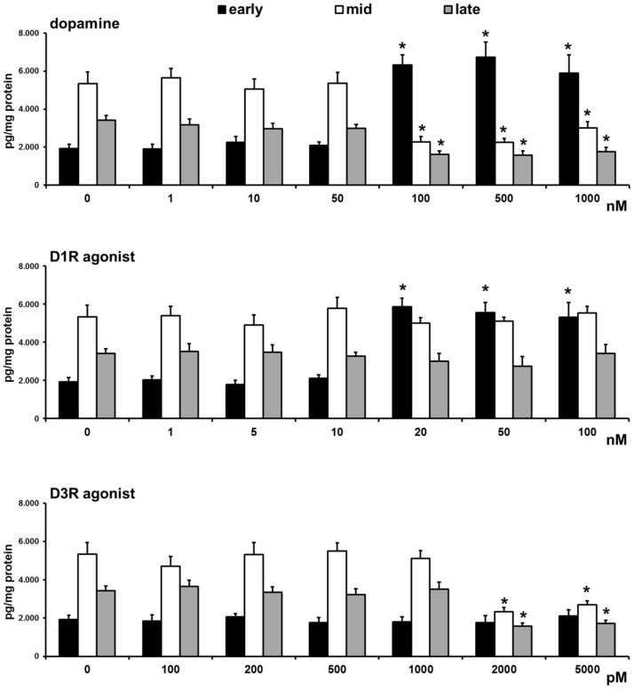
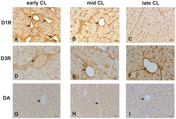
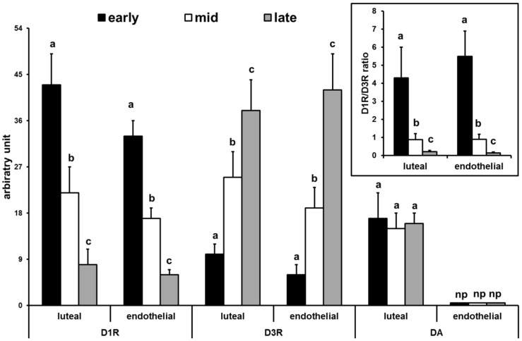
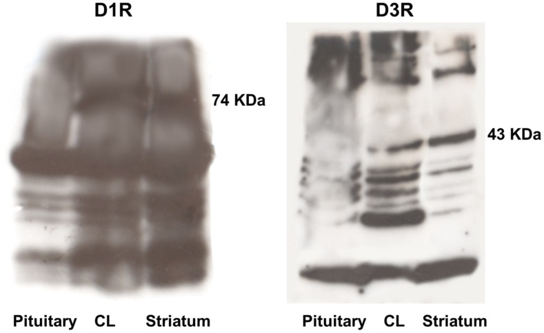
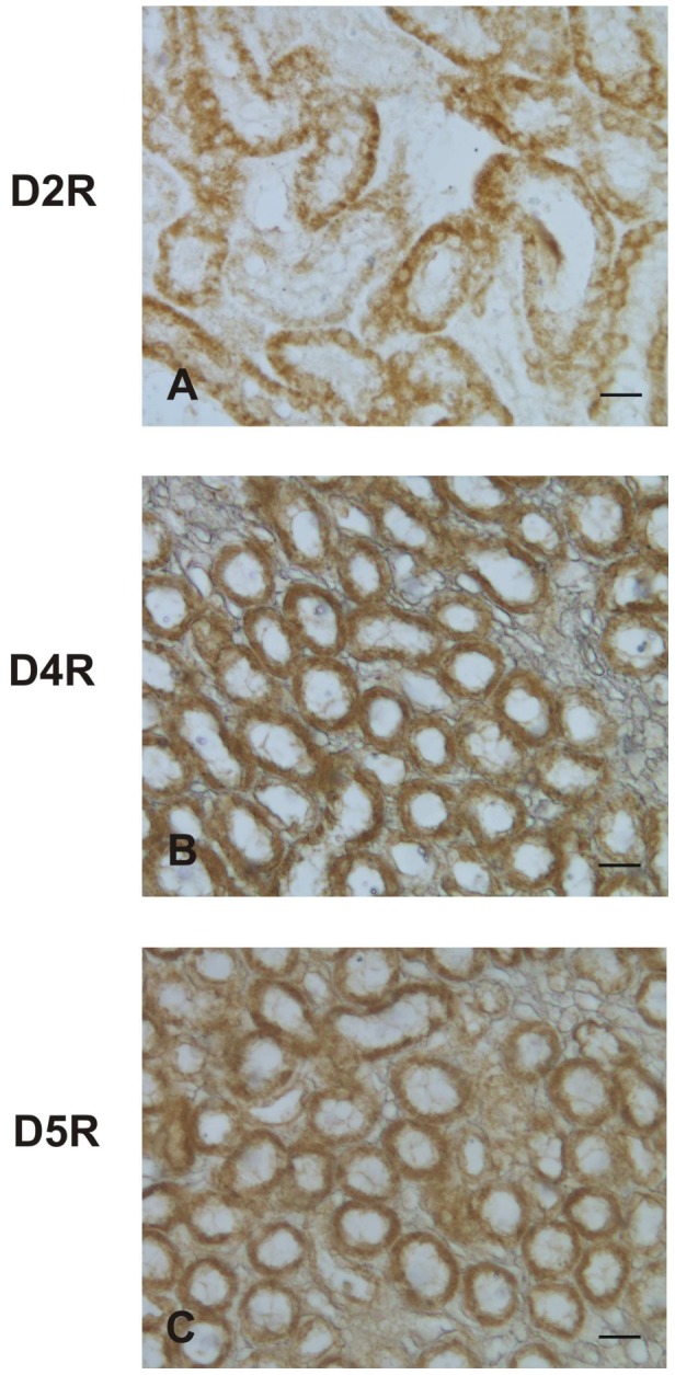
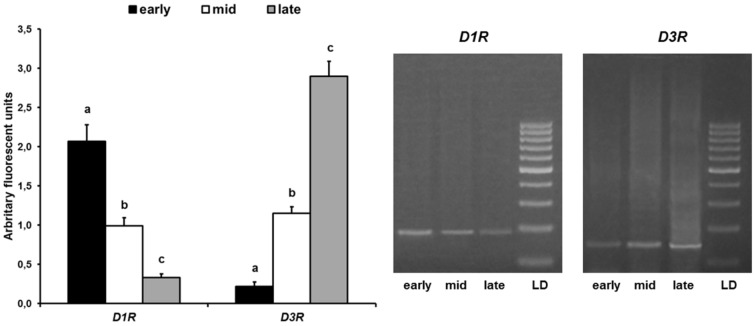
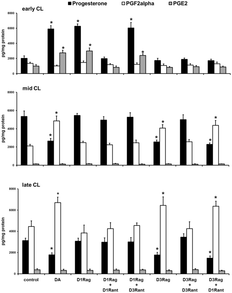
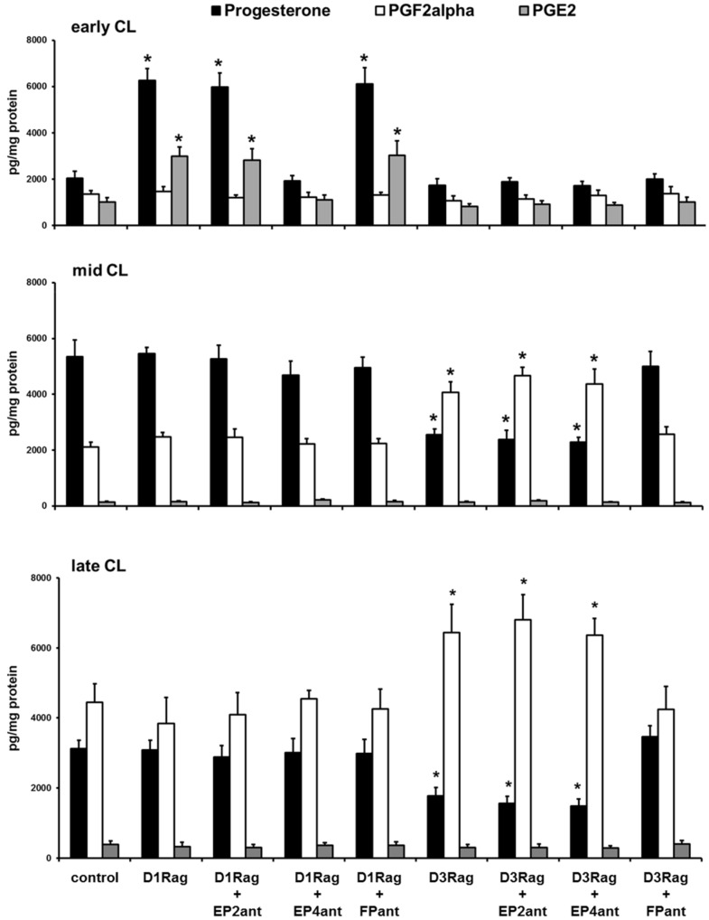
Dopamine (DA) receptor (DR) type 1 (D1R) has been found to be expressed in luteal cells of various species, but the intrinsic role of the DA/DRs system on corpora lutea (CL) function is still unclear. Experiments were devised to characterize the expression of DR types and the presence of DA, as well as the in vitro effects of DA on hormone productions by CL in pseudopregnant rabbits. Immunoreactivity and gene expression for D1R decreased while that for D3R increased in luteal and blood vessel cells from early to late pseudopregnant stages. DA immunopositivity was evidenced only in luteal cells. The DA and D1R agonist increased in vitro release of progesterone and prostaglandin E2 (PGE2) by early CL, whereas the DA and D3R agonist decreased progesterone and increased PGF2α in vitro release by mid- and late CL. These results provide evidence that the DA/DR system exerts a dual modulatory function in the lifespan of CL: the DA/D1R is luteotropic while the DA/D3R is luteolytic. The present data shed new light on the physiological mechanisms regulating luteal activity that might improve our ability to optimize reproductive efficiency in mammal species, including humans.
Conflict of interest statement
Figures
Similar articles
-
Kisspeptin/kisspeptin receptor system in pseudopregnant rabbit corpora lutea: presence and function.Sci Rep. 2019 Mar 25;9(1):5044. doi: 10.1038/s41598-019-41623-1. Sci Rep. 2019. PMID: 30911071 Free PMC article.
-
Evidence for a luteotropic role of peroxisome proliferator-activated receptor gamma: expression and in vitro effects on enzymatic and hormonal activities in corpora lutea of pseudopregnant rabbits.Biol Reprod. 2013 Mar 14;88(3):62. doi: 10.1095/biolreprod.112.107383. Print 2013 Mar. Biol Reprod. 2013. PMID: 23365414
-
Aglepristone (RU534) effects on luteal function of pseudopregnant rabbits: steroid receptors, enzymatic activities, and hormone productions in corpus luteum and uterus.Anim Reprod Sci. 2013 Apr;138(1-2):118-32. doi: 10.1016/j.anireprosci.2013.02.001. Epub 2013 Feb 16. Anim Reprod Sci. 2013. PMID: 23517855
-
Dimerization of dopamine D1 and D3 receptors in the regulation of striatal function.Curr Opin Pharmacol. 2010 Feb;10(1):87-92. doi: 10.1016/j.coph.2009.09.008. Epub 2009 Oct 17. Curr Opin Pharmacol. 2010. PMID: 19837631 Review.
-
Inter-relationships between endothelin and prostaglandin F2alpha in corpus luteum function.Rev Reprod. 2000 Jan;5(1):1-5. doi: 10.1530/ror.0.0050001. Rev Reprod. 2000. PMID: 10711729 Review.
Cited by
-
Antioxidant Intervention Attenuates Aging-Related Changes in the Murine Ovary and Oocyte.Life (Basel). 2020 Oct 22;10(11):250. doi: 10.3390/life10110250. Life (Basel). 2020. PMID: 33105678 Free PMC article.
-
Apelin System in Mammary Gland of Sheep Reared in Semi-Natural Pastures of the Central Apennines.Animals (Basel). 2018 Nov 28;8(12):223. doi: 10.3390/ani8120223. Animals (Basel). 2018. PMID: 30486490 Free PMC article.
-
Increased Serum Levels of Phoenixin-14, Nesfatin-1 and Dopamine Are Associated with Positive Pregnancy Rate after Ovarian Stimulation.J Clin Med. 2023 Nov 8;12(22):6991. doi: 10.3390/jcm12226991. J Clin Med. 2023. PMID: 38002606 Free PMC article.
-
The relationship between polycystic ovary syndrome and gynecological cancers: neurotransmitter metabolism changes and immune regulation.Front Immunol. 2025 Jun 4;16:1578470. doi: 10.3389/fimmu.2025.1578470. eCollection 2025. Front Immunol. 2025. PMID: 40534880 Free PMC article. Review.
-
Kisspeptin/kisspeptin receptor system in pseudopregnant rabbit corpora lutea: presence and function.Sci Rep. 2019 Mar 25;9(1):5044. doi: 10.1038/s41598-019-41623-1. Sci Rep. 2019. PMID: 30911071 Free PMC article.
References
-
- Beaulieu JM, Gainetdinov RR (2011) The physiology, signaling, and pharmacology of dopamine receptors. Pharmacol Rev 63: 182–217. - PubMed
-
- Mayerhofer A, Hemmings HC Jr, Snyder GL, Greengard P, Boddien S, et al. (1999) Functional dopamine-1 receptors and DARPP-32 are expressed in human ovary and granulosa luteal cells in vitro. J Clin Endocrinol Metab 84: 257–264. - PubMed
-
- Mayerhofer A, Fritz S, Grunert R, Sanders SL, Uffy DM, et al. (2000) D1-receptor, darpp-32, and pp-1 in the primate corpus luteum and luteinized granulosa cells: evidence for phosphorylation of darpp-32 by dopamine and human chorionic gonadotropin. J Clin Endocrinol Metab 85: 4750–4757. - PubMed
Publication types
MeSH terms
Substances
LinkOut - more resources
Full Text Sources
Other Literature Sources
