. 2014 Aug 22;9(8):e104989.
doi: 10.1371/journal.pone.0104989. eCollection 2014.

Individual differences in impulsivity predict head motion during magnetic resonance imaging

Affiliations

Individual differences in impulsivity predict head motion during magnetic resonance imaging

Xiang-Zhen Kong et al. PLoS One. .

Abstract

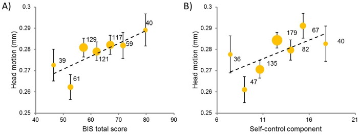
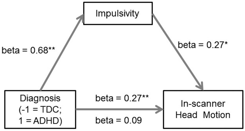
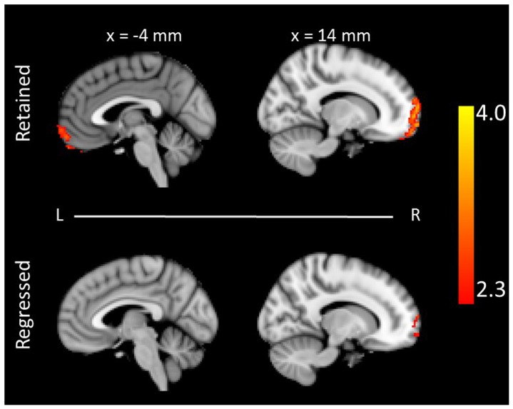
Magnetic resonance imaging (MRI) provides valuable data for understanding the human mind and brain disorders, but in-scanner head motion introduces systematic and spurious biases. For example, differences in MRI measures (e.g., network strength, white matter integrity) between patient and control groups may be due to the differences in their head motion. To determine whether head motion is an important variable in itself, or just simply a confounding variable, we explored individual differences in psychological traits that may predispose some people to move more than others during an MRI scan. In the first two studies, we demonstrated in both children (N = 245) and adults (N = 581) that head motion, estimated from resting-state functional MRI and diffusion tensor imaging, was reliably correlated with impulsivity scores. Further, the difference in head motion between children with attention deficit hyperactivity disorder (ADHD) and typically developing children was largely due to differences in impulsivity. Finally, in the third study, we confirmed the observation that the regression approach, which aims to deal with motion issues by regressing out motion in the group analysis, would underestimate the effect of interest. Taken together, the present findings provide empirical evidence that links in-scanner head motion to psychological traits.


Conflict of interest statement

Competing Interests: The authors have confirmed that Yong He and Yu-Feng Zang are PLOS ONE Editorial Board members, but this does not alter the authors' adherence to PLOS ONE Editorial policies and criteria.

Figures

Figure 1
Figure 1. Binned scatter plots between in-scanner head motion and impulsivity indexed by (A) BIS total score, and (B) self-control component score.
To avoid overlap for participants with similar scores, participants are binned into groups on the basis of impulsivity scores. The size of dots indicates the number of participants in the groups. Error bars indicate standard error of the mean (s.e.m).
Figure 2
Figure 2. Impulsivity as a sufficient mediator of the difference in head motion between ADHD and TDC in the mediation analysis.
Path coefficients are shown next to arrows indicating each link in the analysis. For the group difference in head motion, the value above the arrow indicates the zero-order correlation, and the value below the arrow represents the correlation after controlling the mediator of impulsivity. All values represent standardized betas. * indicates p < 0.01, ** indicates p < 0.001, two-tailed. ADHD: attention deficit hyperactivity disorder; TDC: typically developing children.
Figure 3
Figure 3. The neural correlates of impulsivity in the ADHD children.
The ALFF value of the bilateral orbital frontal cortex and prefrontal cortex was positively correlated with the behavioral measure of impulsivity when the variable of head motion was either retained (top, Z > 2.3, p < 0.05, FWE corrected) or regressed out (bottom, Z > 2.3, uncorrected). The coordinate is in the MNI stereotactic space. ALFF: the amplitude of low-frequency fluctuation.

References

    1. Friston KJ, Williams S, Howard R, Frackowiak RS, Turner R (1996) Movement-related effects in fMRI time-series. Magn Reson Med 35: 346–355. - PubMed
    1. Bullmore ET, Brammer MJ, Rabe-Hesketh S, Curtis VA, Morris RG, et al. (1999) Methods for diagnosis and treatment of stimulus-correlated motion in generic brain activation studies using fMRI. Hum Brain Mapp 7: 38–48. - PMC - PubMed
    1. Jenkinson M, Bannister P, Brady M, Smith S (2002) Improved optimization for the robust and accurate linear registration and motion correction of brain images. Neuroimage 17: 825–841. - PubMed
    1. Tijssen RH, Jansen JF, Backes WH (2009) Assessing and minimizing the effects of noise and motion in clinical DTI at 3 T. Hum Brain Mapp 30: 2641–2655. - PMC - PubMed
    1. Murphy K, Birn RM, Bandettini PA (2013) Resting-state FMRI confounds and cleanup. NeuroImage. - PMC - PubMed

Publication types