

Relationship between ganglion cell layer thickness and estimated retinal ganglion cell counts in the glaucomatous macula
- PMID: 25148790
- PMCID: PMC4252259
- DOI: 10.1016/j.ophtha.2014.06.047
Relationship between ganglion cell layer thickness and estimated retinal ganglion cell counts in the glaucomatous macula
Abstract
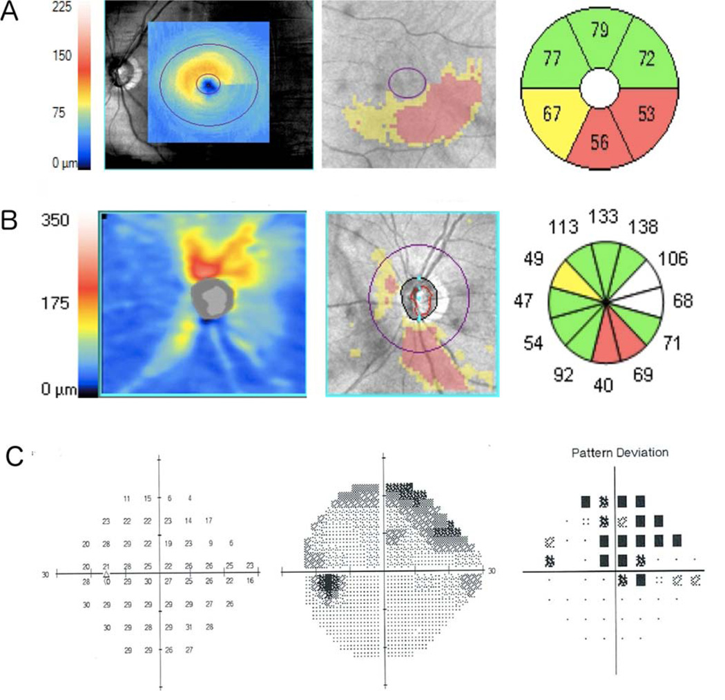
Purpose: To investigate the relationship between macular ganglion cell-inner plexiform layer (mGCIPL) thickness and estimated macular retinal ganglion cell (RGC) counts in glaucoma.
Design: Observational cohort study.
Participants: Cross-sectional study of 77 healthy, 154 glaucoma suspect, and 159 glaucomatous eyes from the Diagnostic Innovations in Glaucoma Study.
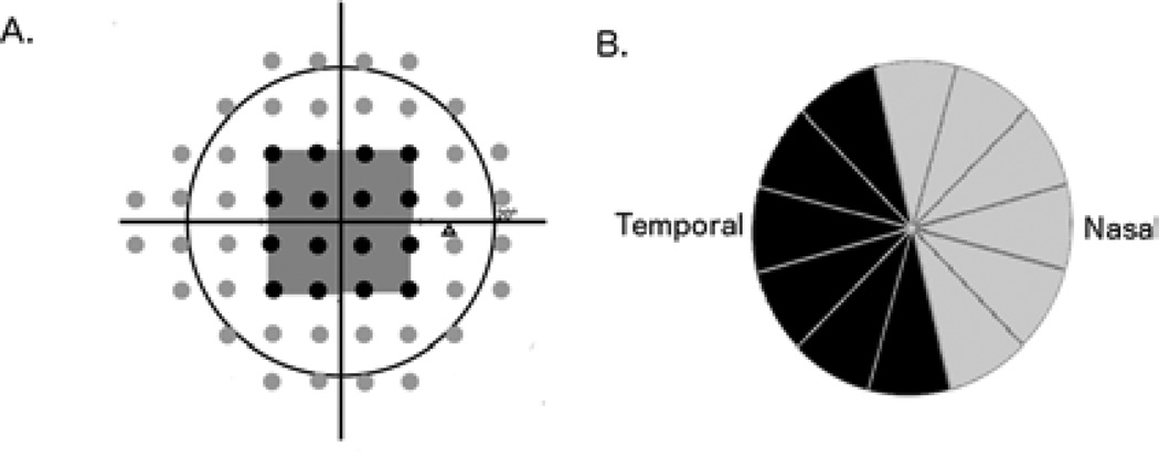
Methods: All eyes underwent 24-2 standard automated perimetry (SAP) and optic nerve and macular imaging using high-definition optical coherence tomography (OCT). The total number of RGCs was estimated using a previously described model that uses SAP and OCT circumpapillary retinal nerve fiber layer (cpRNFL) measurements. The number of macular RGCs was estimated from the temporal cpRNFL and SAP test points within the central 10°.
Main outcome measures: The correlation between mGCIPL thickness and estimates of macular RGC counts.
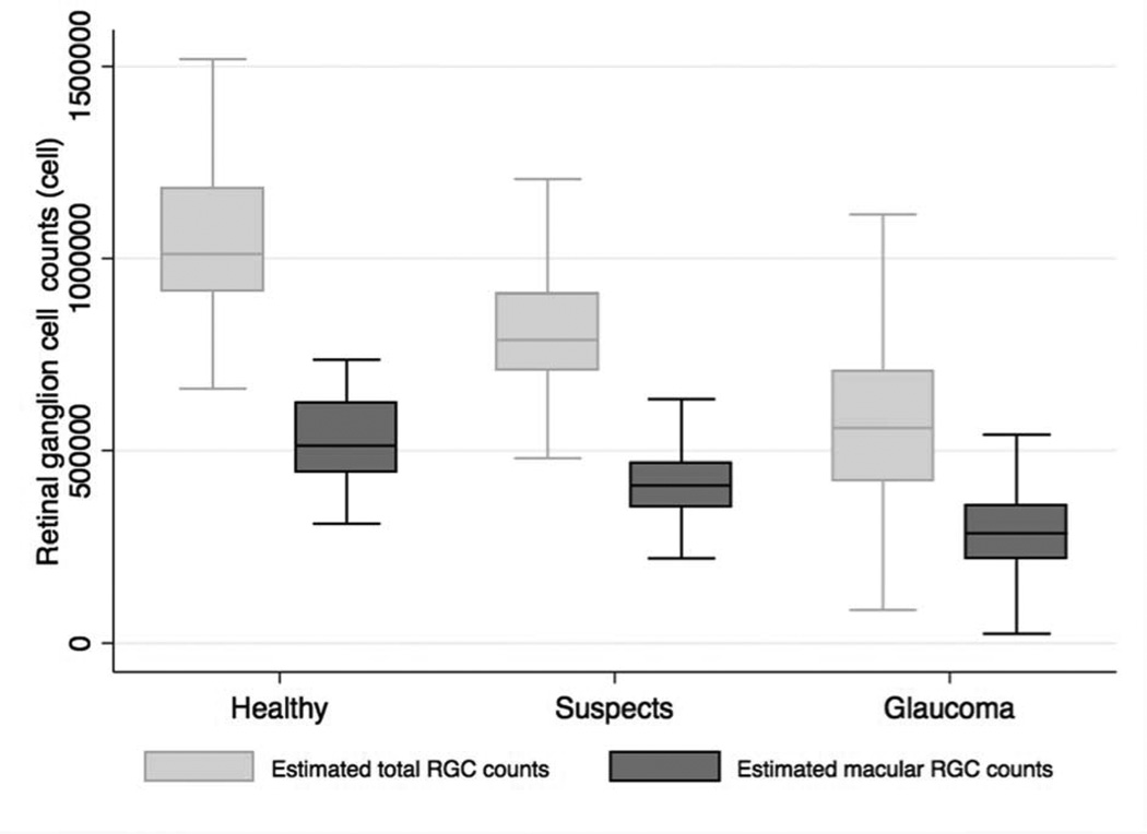
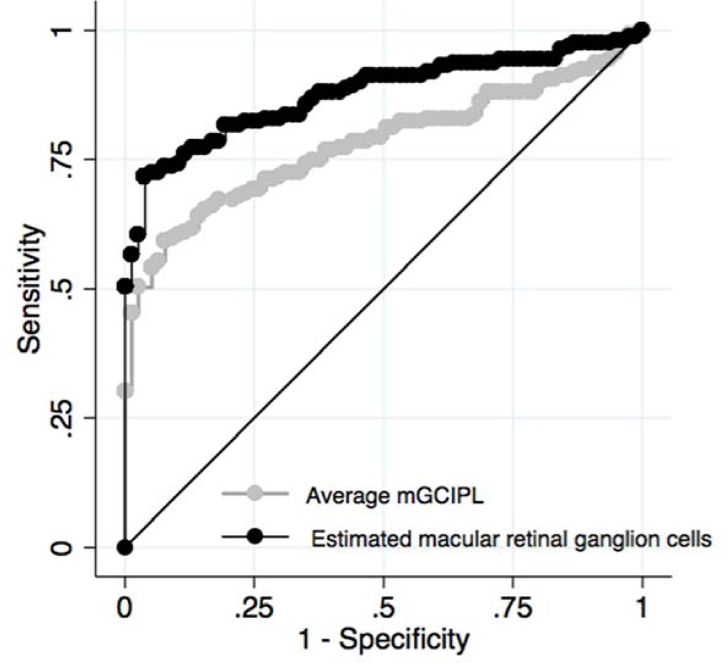
Results: The average estimated macular RGC count in glaucomatous eyes was 306 010 ± 109 449 cells, which was significantly lower than the estimate of 520 678 ± 106 843 cells in healthy eyes (P < 0.001). Glaucomatous eyes had 41% fewer estimated macular RGCs than healthy eyes and suspects had 21% fewer estimated macular RGCs. There was strong correlation between estimated macular RGC counts and mGCIPL thickness (R(2) = 0.67; P < 0.001). Macular RGC counts performed better than average mGCIPL thickness in discriminating healthy and glaucomatous eyes with receiver operating characteristic curve areas of 0.873 and 0.775, respectively (P = 0.015).
Conclusions: The strong association between estimated macular RGC counts and mGCIPL thickness and the better diagnostic performance of the macular RGC counts compared with mGCIPL thickness provides further evidence that estimates of RGC number from cpRNFL thickness and SAP sensitivity can be used to assess neural losses in glaucoma.
Copyright © 2014 American Academy of Ophthalmology. Published by Elsevier Inc. All rights reserved.
Figures

References
-
- Curcio CA, Allen KA. Topography of ganglion cells in human retina. Journal of Comparative Neurology. 1990;300:5–25. - PubMed
-
- Aulhorn E, Karmeyer H. Frequency distribution in early glaucomatous visual field defects. Doc Ophthalmol Proc Ser. 1977;14:75–83.
Publication types
MeSH terms
Grants and funding
LinkOut - more resources
Full Text Sources
Other Literature Sources
Medical
Miscellaneous
