Review
. 2015 Feb;21(1):157-65.
doi: 10.1089/ten.TEB.2014.0383. Epub 2014 Nov 12.

Effect of substrate mechanics on cardiomyocyte maturation and growth

Affiliations
Review

Effect of substrate mechanics on cardiomyocyte maturation and growth

Marwa Tallawi et al. Tissue Eng Part B Rev. 2015 Feb.

Erratum in

Abstract

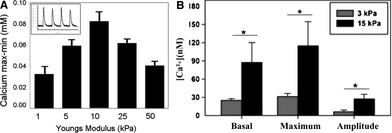
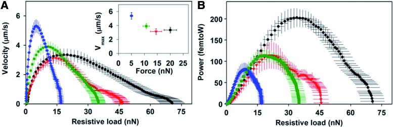
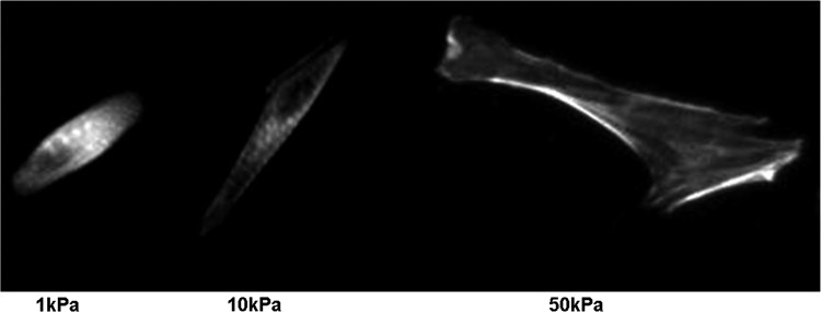
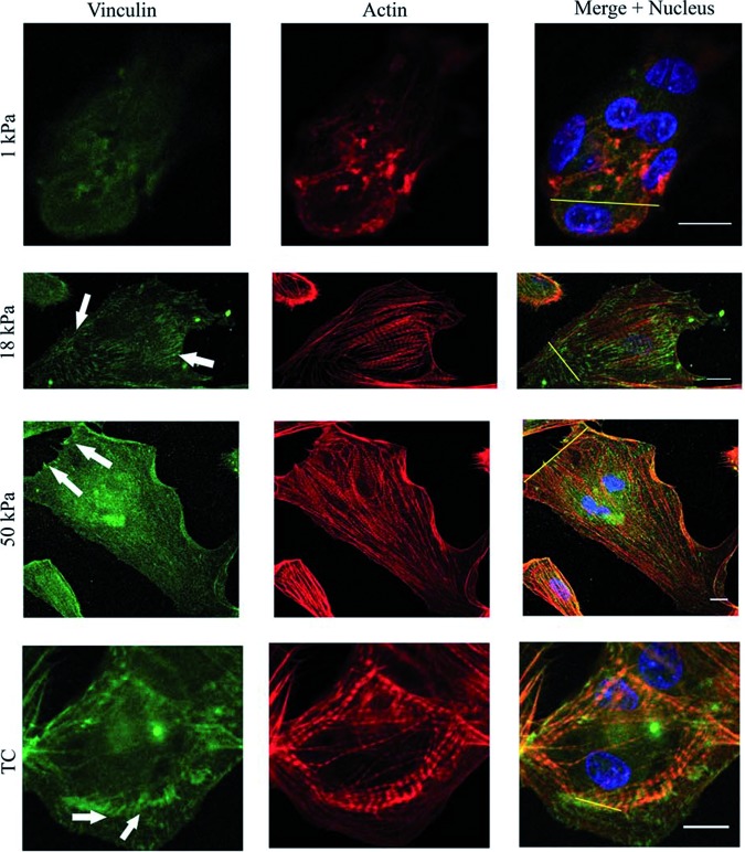
Cardiac tissue engineering constructs are a promising therapeutic treatment for myocardial infarction, which is one of the leading causes of death. In order to further advance the development and regeneration of engineered cardiac tissues using biomaterial platforms, it is important to have a complete overview of the effects that substrates have on cardiomyocyte (CM) morphology and function. This article summarizes recent studies that investigate the effect of mechanical cues on the CM differentiation, maturation, and growth. In these studies, CMs derived from embryos, neonates, and mesenchymal stem cells were seeded on different substrates of various elastic modulus. Measuring the contractile function by force production, work output, and calcium handling, it was seen that cell behavior on substrates was optimized when the substrate stiffness mimicked that of the native tissue. The contractile function reflected changes in the sarcomeric protein confirmation and organization that promoted the contractile ability. The analysis of the literature also revealed that, in addition to matrix stiffness, mechanical stimulation, such as stretching the substrate during cell seeding, also played an important role during cell maturation and tissue development.


Figures

<b>FIG. 1.</b>
FIG. 1.
Micromechanical loading in cardiomyocytes (CMs). (a) The intrinsic load as a result of the contractile activity, (b) an extrinsic load may be applied to stimulate a preload. (c) The microenvironment influences the after-load of tension that is generated in cells in vitro; it can be controlled by surface chemistry, stiffness, topography, and 3D architecture of scaffolds.
<b>FIG. 2.</b>
FIG. 2.
(A) The calcium transients of different matrices was measured as shown in the inset; it is seen that the magnitude of calcium transients on 10 kPa gels was significantly greater than transients on 1 and 50 kPa gels *p<0.05 (reproduced with permission from Liu et al.). (B) The intracellular calcium increases with substrate stiffness. The basal levels, maximum calcium concentration, and the amplitude of the rise in calcium for a twitch contraction increase with substrate stiffness. *p<0.05 (reproduced with permission from Moorman et al.).
<b>FIG. 3.</b>
FIG. 3.
(A) The relationship between twitch force and velocity was plotted for the twitch contraction of CMs cultured on arrays with a stiffness of 3 kPa (blue diamonds), 8 kPa (green squares), 10 kPa (red triangles), and 15 kPa (black circles). Maximum velocity versus the instantaneous force at which maximum velocity was reached; (B) Twitch power was plotted as a function of the resistive load and shows that the maximum power of 200 femtoW increased with substrate stiffness (reproduced with permission from Moorman et al.). Color images available online at www.liebertpub.com/teb
<b>FIG. 4.</b>
FIG. 4.
Neonatal rat ventricular myocytes on collagen-coated polyacrylamide gels and labeled for a α-actinin have poorly defined striations on soft 1 kPa substrates, well-defined and aligned striations on 10 kPa substrates, and unaligned striations with long, large stress fibers on stiff 50 kPa gels (reproduced with permission from Jacot et al.).
<b>FIG. 5.</b>
FIG. 5.
Relative fluorescent intensity of α-actinin and vinculin for cells on the substrates. Sections of cardiac cells expressing vinculin and α-actinin proteins on the different substrates are shown. Fluorescence intensity profiles depict the area of the line drawn in the merged images. The arrows show well-defined mature focal adhesions. Scale bar: 10 μm (reproduced with permission from Bajaj et al.). Color images available online at www.liebertpub.com/teb
<b>FIG. 6.</b>
FIG. 6.
(A) Myocardial elasticity during embryogenesis. Histograms of elastic moduli (∼150 locations per sample) determined from quail embryos show two peaks, one indicating passive elasticity of contractile myocardium (E-Stiff) and a softer, second peak (E-Soft) that surrounds the myocardium and increases in frequency with development. Results for normal and infarcted rat myocardium are indicated for comparison,; (B) CMs and fibroblast spreading. CMs and pericardial fibroblasts were plated from 10-day-old embryonic myocardium without purification, and spread areas were measured after 4 or 24 h5; (C) In vitro striation of CMs. Purified CMs from 10-day-old embryonic myocardium were plated onto substrates of varying elasticity to observe striated cytoskeletal organization with skeletal α-actinin. Many cells on both soft gels and intermediate E* gels reassembled myofibrils, whereas cells on hard matrices exhibited less myofibril reassembly. Inset images show magnified views of the larger images (reproduced with permission from Engler et al.).
<b>FIG. 7.</b>
FIG. 7.
Resting sarcomere length and Z-band width increase with stiffness. (A) Cells on the softest arrays (3 kPa) and (B) on the stiffest arrays (15 kPa) were fixed and stained for α-actinin (green) and nuclei (blue). Insets: Same cell with a bright-field image showing the microposts. (C) Sarcomere length and (D) Z-band width increased with substrate stiffness. Error bars represent the 95% confidence intervals. Scale bars=5 μm (reproduced with permission from Moorman et al.). Color images available online at www.liebertpub.com/teb

References

    1. Roger V.L., Go A.S., Lloyd-Jones D.M., Benjamin E.J., Berry J.D., Borden W.B., et al. . Heart disease and stroke statistics-2012 update: a report from the American Heart Association. Circulation 125,e2, 2012 - PMC - PubMed
    1. Patel J.K., and Kobashigawa J.A.Cardiology patient page. Heart transplantation. Circulation 124,E132, 2011 - PubMed
    1. Jawad H., Lyon A., Harding S.E., Ali N., and Boccaccini A.Myocardial tissue engineering. Br Med Bull 87,31, 2008 - PubMed
    1. Soonpaa M.H., and Field L.J.Survey of studies examining mammalian cardiomyocyte DNA synthesis. Circ Res 83,15, 1998 - PubMed
    1. Engler A., Carag-Krieger C., Johnson C., Raab M., Tang H., Speicher D., et al. . Embryonic cardiomyocytes beat best on a matrix with heart-like elasticity: scar-like rigidity inhibits beating. J Cell Sci 121,3794, 2008 - PMC - PubMed

Publication types