. 2014 Nov:76:55-70.
doi: 10.1016/j.yjmcc.2014.08.009. Epub 2014 Aug 19.

Functional importance of cardiac enhancer-associated noncoding RNAs in heart development and disease

Affiliations

Functional importance of cardiac enhancer-associated noncoding RNAs in heart development and disease

Samir Ounzain et al. J Mol Cell Cardiol. 2014 Nov.

Abstract

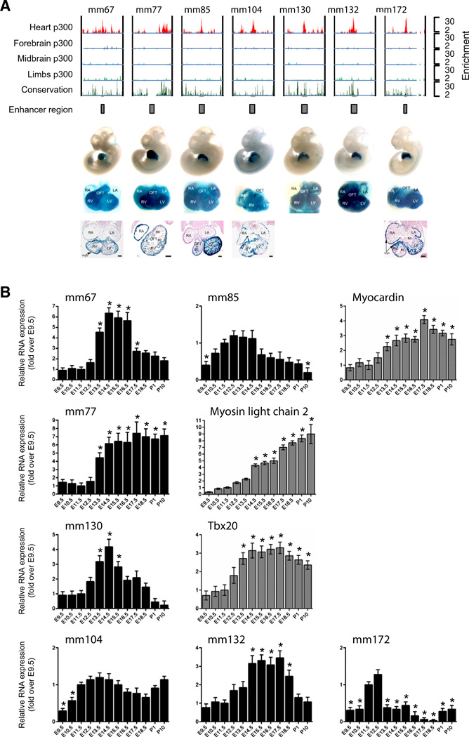
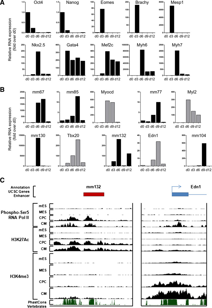
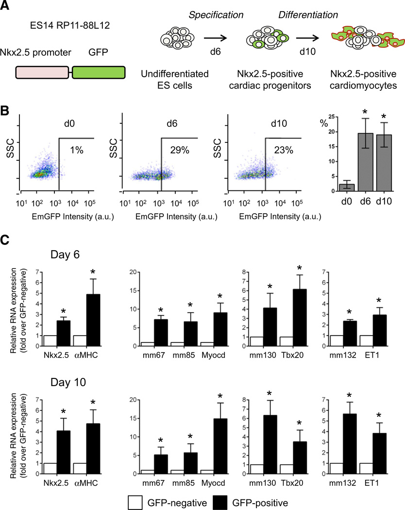
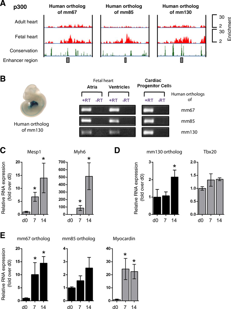
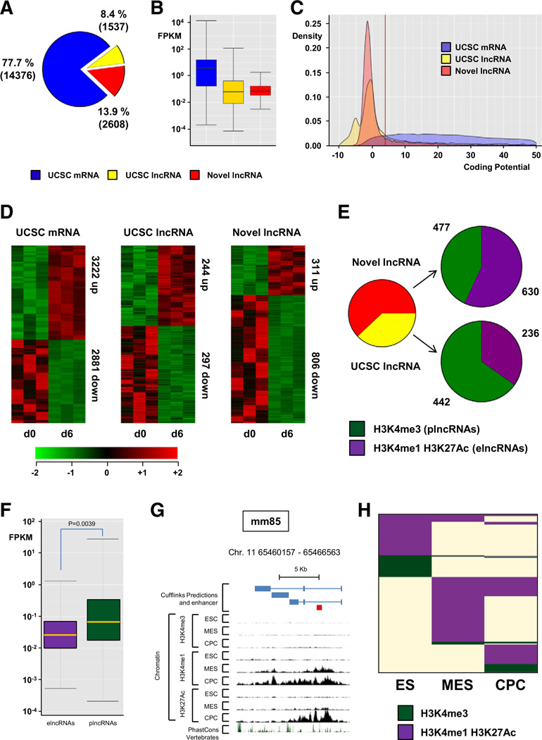
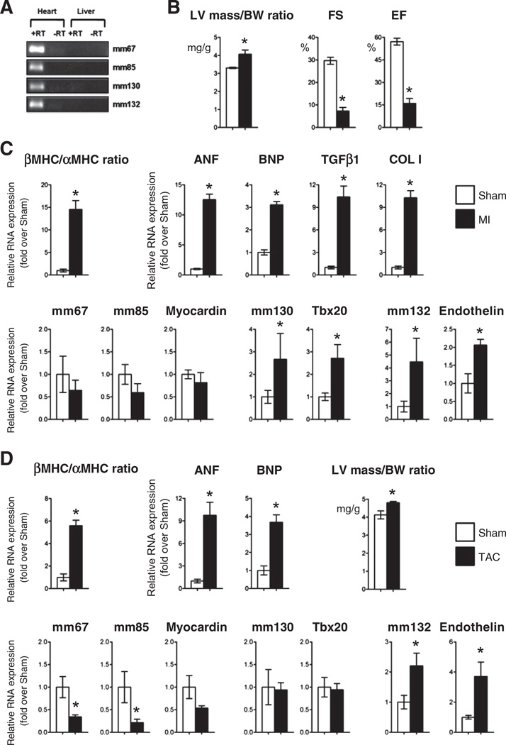
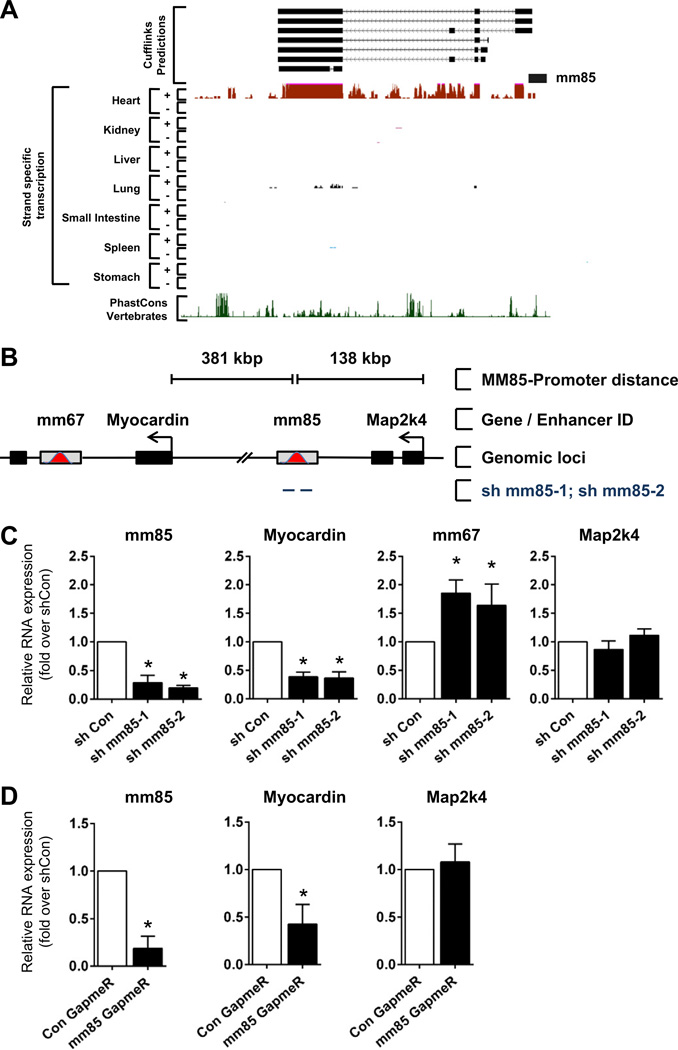
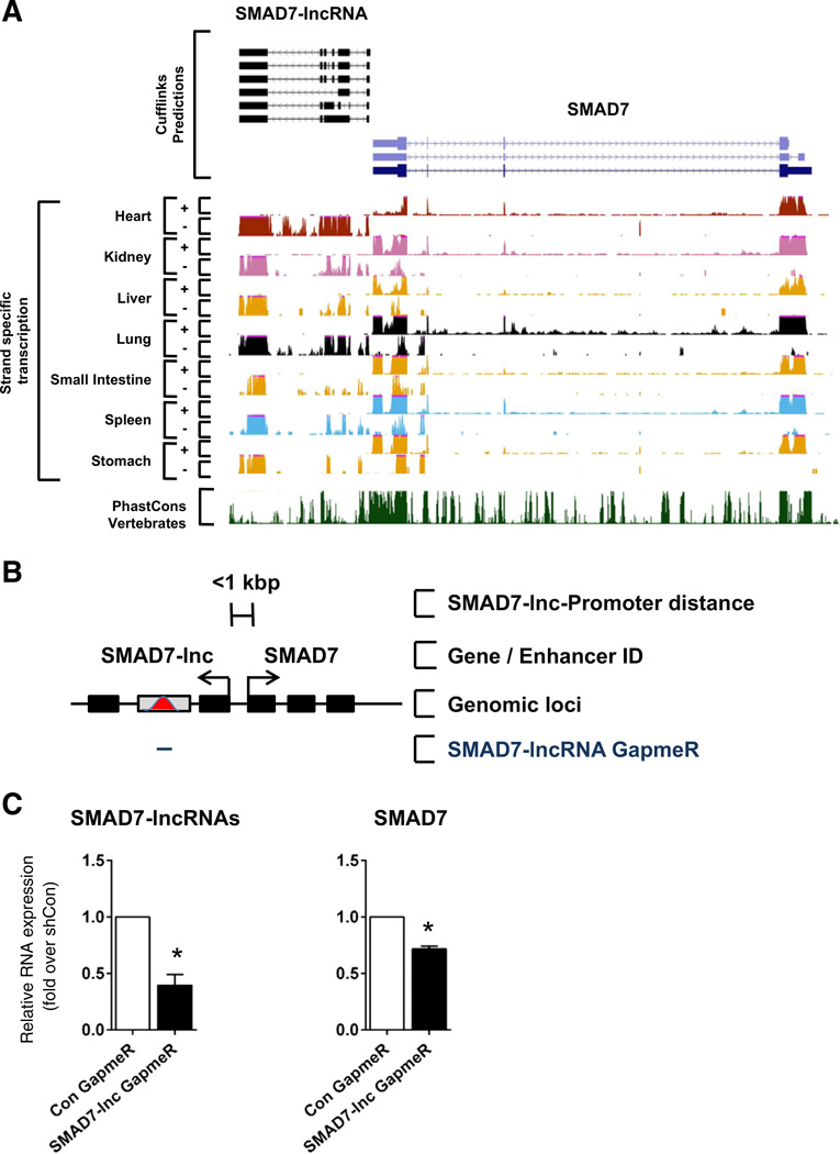
The key information processing units within gene regulatory networks are enhancers. Enhancer activity is associated with the production of tissue-specific noncoding RNAs, yet the existence of such transcripts during cardiac development has not been established. Using an integrated genomic approach, we demonstrate that fetal cardiac enhancers generate long noncoding RNAs (lncRNAs) during cardiac differentiation and morphogenesis. Enhancer expression correlates with the emergence of active enhancer chromatin states, the initiation of RNA polymerase II at enhancer loci and expression of target genes. Orthologous human sequences are also transcribed in fetal human hearts and cardiac progenitor cells. Through a systematic bioinformatic analysis, we identified and characterized, for the first time, a catalog of lncRNAs that are expressed during embryonic stem cell differentiation into cardiomyocytes and associated with active cardiac enhancer sequences. RNA-sequencing demonstrates that many of these transcripts are polyadenylated, multi-exonic long noncoding RNAs. Moreover, knockdown of two enhancer-associated lncRNAs resulted in the specific downregulation of their predicted target genes. Interestingly, the reactivation of the fetal gene program, a hallmark of the stress response in the adult heart, is accompanied by increased expression of fetal cardiac enhancer transcripts. Altogether, these findings demonstrate that the activity of cardiac enhancers and expression of their target genes are associated with the production of enhancer-derived lncRNAs.

Keywords: Cardiac development; Enhancers; Gene regulation; Gene regulatory networks; Heart failure; Long noncoding RNA (lncRNAs).


Figures

Fig. 1
Fig. 1
Fetal cardiac enhancers are expressed in the developing heart. A. ChIP-Seq profiles of p300 occupancy at cardiac enhancers. Coverage by extended p300 reads in heart (red), forebrain (dark blue), midbrain (light blue) and limb (green). Vertebrate conservation plots (black) were obtained from the UCSC genome browser. Gray boxes correspond to candidate enhancer region. Numbers at the right indicate overlapping extended reads. Below boxes are LacZ-stained embryos and isolated hearts with in vivo enhancer activity at E11.5. B. Total RNA was extracted from embryonic (E9.5–E18.5) and neonatal (P1–P10) mouse hearts and subjected to reverse transcription followed by quantitative RT-PCR. Enhancer-associated transcripts: mm67, mm85, mm77, mm104, mml 30, mm132 and mm172, and their putative target genes: Myocardin, Myosin light chain 2 and Tbx20. Results are normalized for expression in the E11.5 heart Mean ± SEM; n = 6–8. * indicates statistical significance, p < 0.05.
Fig. 2
Fig. 2
Fetal cardiac enhancers are expressed during embryonic stem cell differentiation into the cardiogenic lineage. RNA was isolated on d0, d3, d6, d9 and d12 of embryoid body formation. A. Relative RNA levels of stage-specific markers of cardiac differentiation. B. Relative RNA levels of enhancer-associated ncRNAs and putative target genes during cardiac differentiation. C. UCSC genome browser views of ChIP-Seq data at the mm132 and Edn1 loci for mES, MES, CPC and CM representing different stages of cardiac differentiation.
Fig. 3
Fig. 3
Fetal cardiac enhancer-derived transcripts are enriched in Nkx2.5-positive cardiac progenitor cells. A. Schematic of ES cell differentiation, and isolation of Nkx2.5-positive cardiac progenitors and cardiomyocytes. B. Time course of EmGFP expression during embryoid-body formation and cardiac differentiation at d0, d6 and d10. Bars indicate mean percentages of EmGFP (Nkx2.5)-positive cells at d0, d6 and d10 of differentiation (n = 3); C. Relative RNA levels of enhancer-associated ncRNAs and putative target genes in sorted EmGFP (Nkx2.5)-positive (black bar) and EmGFP (Nkx2.5)-negative (white bar) cells. Mean ± SEM; n = 3. * indicates statistical significance, p < 0.05.
Fig. 4
Fig. 4
Orthologous human enhancer sequences are expressed in the fetal heart and in differentiating cardiac progenitor cells. A. ChIP-Seq profiles of p300/CBP occupancy in genomic regions of human orthologous enhancer sequences are indicated by red peaks. Black boxes in the lower panel correspond to the enhancer sequence. Vertebrate conservation plots (black) were obtained from the UCSC browser. B. In vivo activity of human orthologous mml30 enhancer in E11.5 transgenic mice (left panel). Fetal cardiac enhancers are expressed in both cardiac chambers and isolated cardiac progenitor cells (CPCs)(right panel); C. Relative RNA levels of cardiac differentiation markers in differentiating human CPCs; D and E. Relative RNA levels of enhancer-associated ncRNAs and putative target genes during cardiac differentiation of human CPCs. RNA was isolated on d0, d7 and d14 of differentiation. Mean ± SEM; n = 4. * indicates statistical significance, p < 0.05.
Fig. 5
Fig. 5
Global discovery of enhancer associated IncRNA expression in differentiating mESCs. RNA-Seq was performed on RNA samples isolated from undifferentiated mESC (d0) and from differentiating mESc at the cardiac precursor stage (d6 after induction of differentiation) to characterise the differentiation-associated transcriptome. (A) Pie chart showing composition of the Poly(A)+ transcriptome, UCSC mRNAs (blue), UCSC IncRNAs (yellow) and novel IncRNAs (red). (B) Box plot of transcript abundance (fragments per kilobase per million reads [FPKM]) of UCSC mRNAs (blue), UCSC IncRNAs (yellow) and novel IncRNAs (red). (C) Kernel density plot of coding potential (Gene ID score). (D) Heat maps showing hierarchical clustering of differentially expressed transcripts within the three RNA classes during ESC differentiation. (E) Pie charts showing distribution of USCS IncRNAs (yellow) and novel IncRNAs (red) associated with a canonical promoter signature (H3K3me3, green) or active enhancer state (H3K4mel/H3K27Ac) during ESC differentiation. (F) Box plot of transcript abundance (FPKM) of enhancer-templated (purple) and canonical promoter-associated (green) IncRNAs. (G) The mm85-templated IncRNA is associated with acquisition of active enhancer state during ESC differentiation. (H) Chromatin state map of all IncRNAs associated with either canonical promoter or enhancer state in at least one lineage, ES cell (ES), mesodermal precursors (MES) and cardiac precursor cells (CPCs). Rows are recursively clustered by these marks in these lineages.
Fig. 6
Fig. 6
Fetal enhancer expression is induced in response to stress in the adult heart A. Fetal enhancers are expressed in the adult mouse heart B. Cardiac dimensions and function in mice 14 days after myocardial infarction. C. Relative RNA levels of cardiac stress markers, enhancer-associated ncRNAs and target genes in sham-operated (Sham; white bar) and myocardial infarction (M; black bar) groups. Ratio of β over α Myosin heavy chain expression is also indicated. Results are normalized to levels measured in sham-operated mice. Mean ± SEM; n = 4–6. * indicates statistical significance, p < 0.05. D. Left ventricular mass to body weight ratio, relative RNA levels of cardiac stress markers, mm132 enhancer-associated ncRNAs and Endothelin in sham-operated (Sham; white bar) and transaortic constriction (TAC; black bar) groups. Ratio of β over α Myosin heavy chain expression is also indicated. Results are normalized to levels measured in sham-operated mice. Mean ± SEM; n = 3–5. * indicates statistical significance, p < 0.05.
Fig. 7
Fig. 7
The IncRNA associated to the mm85 fetal enhancer is required for transcription of its target gene. A. UCSC genome browser views of strand-specific RNA-Seq data at mm85 genomic locus for adult heart, kidney, liver, lung, small intestine, spleen and stomach. B. Schematic illustrating the relative genomic location and distance of fetal mm85 enhancer, fetal enhancer mm67, Myocardin proximal target genes and Map2k4 gene. Black bars indicate protein coding genes, gray bars with red peak indicate fetal enhancers. Tailed lines describe relative distances between mm85 and proximal gene transcription start sites. C. P19CL6 cells were transfected with the indicated shRNAs directed against mm85. Relative levels of mm85, Myocardin, mm67 and Map2k4 RNAs are normalized to levels found in cells transfected with control shRNA (shCon). Mean ± SEM; n = 3. * indicates statistical significance, p < 0.05. D. Mouse neonatal CMs were transfected with GapmeRs targeting mm85 IncRNA or random scrambled sequence. Cells were harvested 48 h post transfection and assayed for mm85 IncRNA, Myocardin and Map2k4 expression by qRT-PCR. Bars represent mean ± SEM; n = 2. * indicates statistical significance, p < 0.05.
Fig. 8
Fig. 8
The SMAD7-associated enhancer-derived IncRNA is required for its transcription. A. UCSC genome browser views of strand-specific RN A-Seq data at SMAD7 genomic locus for adult heart, kidney, liver, lung, small intestine, spleen and stomach. B. Schematic illustrating the relative genomic location and distance of the SMAD7-lncRNA and SMAD7. Black bars indicate coding and noncoding exons, gray bars with red peak indicate fetal enhancers. Tailed lines describe relative distances between IncRNA and SMAD7 transcriptional start sites. C. Neonatal cardiac fibroblasts were transfected with GapmeRs targeting SMAD7-lncRNA or random scrambled sequence. Cells were harvested 48 h post transfection and assayed for SMAD7-lncRNA and SMAD7 expression by qRT-PCR Bars represent mean ± SEM; n = 4. * indicates statistical significance, p < 0.05.

Comment in

  • Non-coding RNA enhances cardiac development.
    Hofmann P, Boon RA. Hofmann P, et al. J Mol Cell Cardiol. 2014 Nov;76:205-7. doi: 10.1016/j.yjmcc.2014.09.005. Epub 2014 Sep 18. J Mol Cell Cardiol. 2014. PMID: 25240640 No abstract available.

Similar articles

Cited by

References

    1. Olson EN. Gene regulatory networks in the evolution and development of the heart. Science. 2006;313(5795):1922–1927. - PMC - PubMed
    1. Bruneau BG. Transcriptional regulation of vertebrate cardiac morphogenesis. Circ Res. 2002;90(5):509–519. - PubMed
    1. Bruneau BG. The developmental genetics of congenital heart disease. Nature. 2008;451(7181):943–948. - PubMed
    1. Ong CT, Corces VG. Enhancer function: new insights into the regulation of tissue-specific gene expression. Nat Rev Genet. 2011;12(4):283–293. - PMC - PubMed
    1. Wamstad JA, Alexander JM, Truty RM, Shrikumar A, Li F, Eilertson KE, et al. Dynamic and coordinated epigenetic regulation of developmental transitions in the cardiac lineage. Cell. 2012;151(1):206–220. - PMC - PubMed

Publication types