. 2015 Jan;13(1):149-60.
doi: 10.1158/1541-7786.MCR-14-0110. Epub 2014 Aug 22.

Phosphodiesterase 4D inhibitors limit prostate cancer growth potential

Affiliations

Phosphodiesterase 4D inhibitors limit prostate cancer growth potential

Ginny L Powers et al. Mol Cancer Res. 2015 Jan.

Abstract

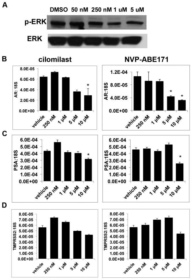
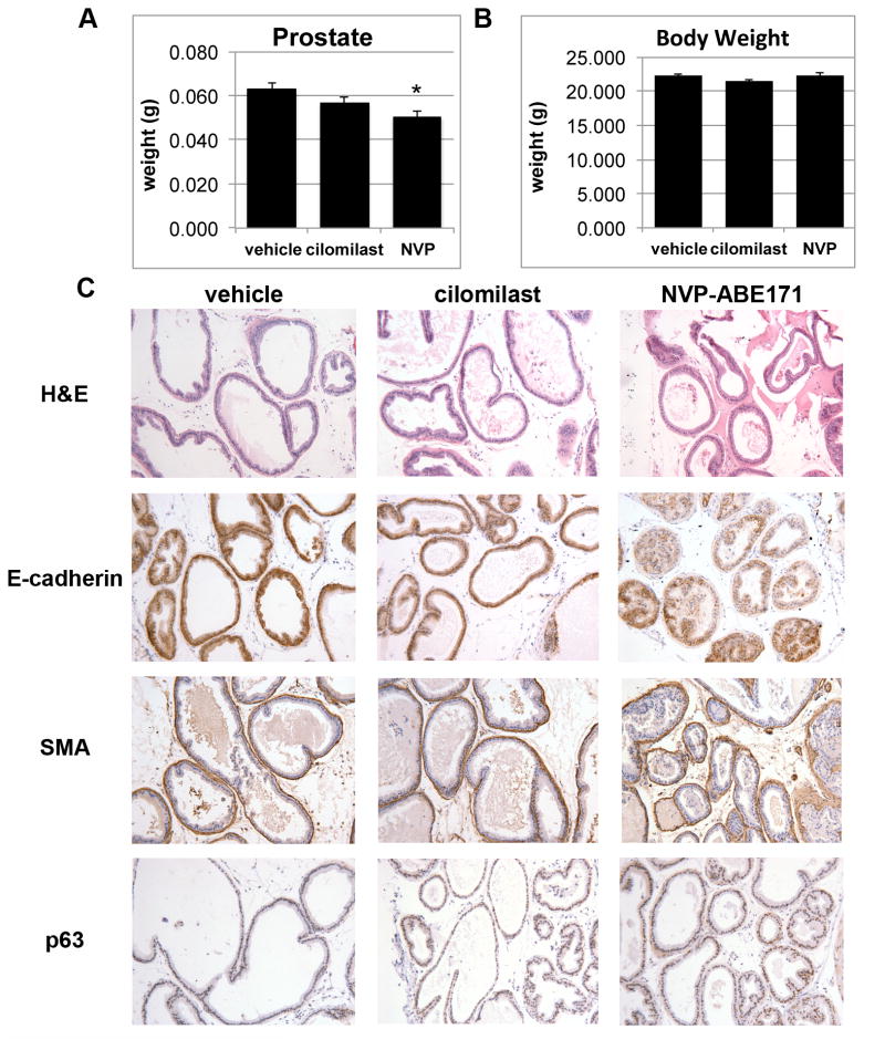
Phosphodiesterase 4D (PDE4D) has recently been implicated as a proliferation-promoting factor in prostate cancer and is overexpressed in human prostate carcinoma. However, the effects of PDE4D inhibition using pharmacologic inhibitors have not been examined in prostate cancer. These studies examined the effects of selective PDE4D inhibitors, NVP-ABE171 and cilomilast, as anti-prostate cancer therapies in both in vitro and in vivo models. The effects of PDE4D inhibitors on pathways that are critical in prostate cancer and/or downstream of cyclic AMP (cAMP) were examined. Both NVP-ABE171 and cilomilast decreased cell growth. In vitro, PDE4D inhibitors lead to decreased signaling of the sonic hedgehog (SHH), androgen receptor (AR), and MAPK pathways, but growth inhibition was best correlated to the SHH pathway. PDE4D inhibition also reduced proliferation of epithelial cells induced by paracrine signaling from cocultured stromal cells that had activated hedgehog signaling. In addition, PDE4D inhibitors decreased the weight of the prostate in wild-type mice. Prostate cancer xenografts grown in nude mice that were treated with cilomilast or NVP-ABE171 had decreased wet weight and increased apoptosis compared with vehicle-treated controls. These studies suggest the pharmacologic inhibition of PDE4D using small-molecule inhibitors is an effective option for prostate cancer therapy.

Implications: PDE4D inhibitors decrease the growth of prostate cancer cells in vivo and in vitro, and PDE4D inhibition has therapeutic potential in prostate cancer.


Conflict of interest statement

Conflict of Interest: The authors declare no conflict of interest.

Figures

Figure 1
Figure 1. LNCaP-C4 cells have decreased growth when treated with PDE4D inhibitors cilomilast and NVP-ABE171
LNCaP-C4 prostate cancer cells in reduced serum media were treated with PDE4D selective inhibitors for 72 hours with A) cilomilast or B) NVP-ABE171 or vehicle-only (DMSO) control. BPH1 benign prostatic hyperplasia cells were treated with vehicle control (DMSO), C) cilomilast or D) NVP-ABE171 in complete media. E) P2 cells were treated with the indicated dose of NVP-ABE171 for 72 hours in reduced serum media. F) BHPrE1 or G) NHPrE1 cells were treated with 5 μM cilomilast or 50 nM NVP-ABE171 for 72 hours in reduced serum media. The graphs depict relative cell numbers for the different treatment groups with data normalized to vehicle-only controls and are presented as percent of vehicle control. Statistically significant differences from the vehicle-only controls were determined by ANOVA followed by Bonferroni's test for multiple comparisons and are indicated on the graph with *P<0.05. Bars indicate the standard error.
Figure 2
Figure 2. PDE4D inhibition affects PKA mediated pathways
LNCaP-C4 cells were treated with vehicle-only control (DMSO), cilomilast, or NVP-ABE171 for 72 hours in reduced serum media. A) Representative Western blot from whole cell lysates of LNCaP-C4 cells treated with indicated doses of NVP-ABE171.B-D) RNA was isolated, reverse transcribed, and qRT-PCR was performed to evaluate gene expression. Expression of B) Androgen Receptor and its target genes C) PSA and D) TMPRSS2 were assessed with cilomilast and NVP-ABE171 treatment. Data are presented as a representative experiment selected from a minimum of three independent experiments. Bars represent the average of three replicates and error bars are the SEM. ANOVA with Tukey's post test was performed and statistical significance (p<0.05) compared with vehicle control is indicated by an *.
Figure 3
Figure 3. PDE4D inhibition decreased autocrine hedgehog signaling
LNCaP-C4 cells in reduced serum media were treated with vehicle-only control (DMSO), or the indicated concentrations of cilomilast, or NVP-ABE171 for 72 hours and RNA was collected. A) PTCH1, B) GLI1, and 18S transcript levels were evaluated using qRT-PCR. Statistical analysis was evaluated with ANOVA using GraphPad Prism. *P<0.05, (n=3 per data point). In conditions where LNCaP-C4 cell proliferation was reduced by NVP-ABE171 and cilomilast, Ptch1 and Gli1 mRNA expression was reduced. C) LNCaP-C4 cells were treated with vehicle-only control, 25 nM NVP-ABE171 (NVP), 0.5 μM cyclopamine (cyclo), or the combination of 25 nM NVP-ABE171 and 0.5 μM cyclopamine in reduced serum media. Cell growth was assessed after 72 hours of treatment. Statistically significant differences were determined by ANOVA followed by Bonferroni's test for multiple comparisons and are indicated on the graph with *P<0.05 (n=6 per data point). Bars indicate the standard error.
Figure 4
Figure 4. Inhibition of PDE4D reduced growth and affected gene expression induced by Shh signaling in epithelial and stromal cells
A) LNCaP or B) LNCaP-C4 cells were cultured in a microculture device with either UGSM-2, UGSM-2 + Shh, or Gli3-/- UGSM cells. Drugs (vehicle, cilomilast, or NVP-ABE171) were added at day 1 and replenished until day 5. Proliferation was assessed by EdU. Statistically significant differences were determined by ANOVA followed by Bonferroni's test for multiple comparisons and are indicated on the graph with *P<0.05 (n=6 per data point). Bars indicate the standard error. C-D) LNCaP cells were cultured in a microculture device with either UGSM-2, UGSM-2 + Shh, or Gli3-/- UGSM cells. Drugs were added at day 1 and replenished until day 5. RNA was extracted from the mesenchymal cells and C) Gli1 and D) Ptch1 expression was examined. Statistically significant differences were determined by ANOVA followed by Bonferroni's test for multiple comparisons and are indicated on the graph with *P<0.05 (n=6 per data point, relative to appropriate vehicle control). Error bars indicate the standard error.
Figure 5
Figure 5. PDE4D inhibition decreased prostate weight but did not alter prostate morphology
A-B) Male C57BL/6 mice were treated with vehicle-only (10% EtOH, 90% olive oil) vehicle (v), cilomilast (c) or NVP-ABE171 (N) by oral gavage daily (n=5-6 per condition). After six weeks animals were sacrificed and prostate were collected. Average weights of the A) prostate and B) total animal are represented in the graphs and error bars represent the SEM. Statistical analysis was performed using an ANOVA followed by Tukey's test for individual comparisons (* NVP-ABE171 P<0.05 versus vehicle control). C) IHC was performed on prostates that were fixed and paraffin sectioned. Sections were stained by H&E to assess overall morphology and IHC was performed to examine epithelial (E-cadherin), stromal (SMA- smooth muscle actin), and basal cell (p63) markers.
Figure 6
Figure 6. PDE4D inhibitors increased apoptosis and decreased proliferation of LNCaP-C4 xenografts
LNCaP-C4 prostate cancer cells were suspended in matrigel and transplanted under the kidney capsule of male nude mice hosts. Mice were treated with vehicle-only (10% EtOH, 90% olive oil) control, cilomilast or NVP-ABE171 by gavage (n=7-11 for each respective group). A) After 6 weeks of treatment, the xenografts were excised, weighed, and photographed. B) The average wet weights of the xenografts are shown with bars indicating the standard error. Statistical analysis was determined by ANOVA followed by Tukey's test for individual comparisons, *P<0.05 for cilomilast, P<0.05 for NVP-ABE171 versus vehicle control. C) The xenografts LNCaP-C4 xenografts were excised, fixed in formalin, and embedded in paraffin blocks. IHC with Ki67 (proliferation), TUNEL (apoptosis), or p21 (senescence) was performed. Representative Ki67 and TUNEL photomicrographs for each treatment group are shown. D) Labeling index was assessed for Ki67, TUNEL, and p21. Statistical analysis was determined by ANOVA followed by Tukey's test for individual comparisons, *P<0.05 for NVP-ABE171 versus vehicle-only control for Ki67. *P<0.01 for cilomilast, P<0.001 for NVP-ABE171 versus vehicle-only control for TUNEL.

References

    1. Jemal A, Siegel R, Ward E, Murray T, Xu J, Smigal C, et al. Cancer statistics, 2006. CA: a cancer journal for clinicians. 2006;56:106–30. - PubMed
    1. Rahrmann EP, Collier LS, Knutson TP, Doyal ME, Kuslak SL, Green LE, et al. Identification of PDE4D as a proliferation promoting factor in prostate cancer using a Sleeping Beauty transposon-based somatic mutagenesis screen. Cancer Res. 2009;69:4388–97. - PMC - PubMed
    1. Baca SC, Prandi D, Lawrence MS, Mosquera JM, Romanel A, Drier Y, et al. Punctuated evolution of prostate cancer genomes. Cell. 2013;153:666–77. - PMC - PubMed
    1. Natrajan R, Mackay A, Lambros MB, Weigelt B, Wilkerson PM, Manie E, et al. A whole-genome massively parallel sequencing analysis of BRCA1 mutant oestrogen receptor-negative and -positive breast cancers. J Pathol. 2012;227:29–41. - PMC - PubMed
    1. Houslay MD. PDE4 cAMP-specific phosphodiesterases. Progress in nucleic acid research and molecular biology. 2001;69:249–315. - PubMed

Publication types

MeSH terms