. 2014 Nov;80(21):6694-703.
doi: 10.1128/AEM.01789-14. Epub 2014 Aug 22.

Combined use of bacteriophage K and a novel bacteriophage to reduce Staphylococcus aureus biofilm formation

Affiliations

Combined use of bacteriophage K and a novel bacteriophage to reduce Staphylococcus aureus biofilm formation

D R Alves et al. Appl Environ Microbiol. 2014 Nov.

Abstract

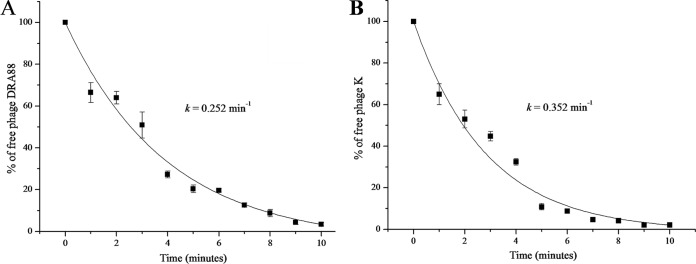
Biofilms are major causes of impairment of wound healing and patient morbidity. One of the most common and aggressive wound pathogens is Staphylococcus aureus, displaying a large repertoire of virulence factors and commonly reduced susceptibility to antibiotics, such as the spread of methicillin-resistant S. aureus (MRSA). Bacteriophages are obligate parasites of bacteria. They multiply intracellularly and lyse their bacterial host, releasing their progeny. We isolated a novel phage, DRA88, which has a broad host range among S. aureus bacteria. Morphologically, the phage belongs to the Myoviridae family and comprises a large double-stranded DNA (dsDNA) genome of 141,907 bp. DRA88 was mixed with phage K to produce a high-titer mixture that showed strong lytic activity against a wide range of S. aureus isolates, including representatives of the major international MRSA clones and coagulase-negative Staphylococcus. Its efficacy was assessed both in planktonic cultures and when treating established biofilms produced by three different biofilm-producing S. aureus isolates. A significant reduction of biofilm biomass over 48 h of treatment was recorded in all cases. The phage mixture may form the basis of an effective treatment for infections caused by S. aureus biofilms.


Figures

FIG 1
FIG 1
Curve for one-step growth of phage DRA88 (left) and phage K (right) in S. aureus RN4220 at 37°C. Shown are the PFU per infected cell in untreated cultures (●) and in chloroform-treated cultures (○) at several time points over 60 min. The phage growth parameters are indicated in the figure and correspond to eclipse period (E), latent period (L), and burst size (B). Each data point is the mean of three independent experiments, and error bars indicate the means ± standard deviations.
FIG 2
FIG 2
Percentage of free DRA88 (A) and phage K (B) phages after infection of actively growing S. aureus RN4220 at an MOI of 0.001 at several time points over 10 min. Rate constants for loss of phage are 0.352 min−1 for phage K and 0.252 min−1 for DRA88. Each data point is the mean of three independent experiments, and error bars indicate the means ± standard deviations.
FIG 3
FIG 3
Electron micrograph images of phage DRA88 negatively stained with 1% uranyl acetate, showing the tail in a contracted position (A) and formation on phage particle aggregates (B). Scale is indicated by the bars.
FIG 4
FIG 4
Comparative genomic analysis of phage DRA88 and phage K. Nucleotide sequences were compared using the Artemis comparison tool (ACT). Predicted open reading frames are denoted by arrows, tRNAs are indicated (vertical blue dashed line), and genes encoding proteins with at least 69% amino acid identity between the two genomes are indicated by shaded regions.
FIG 5
FIG 5
Dynamic of bacteria with single phage and phage mixture in liquid cultures over 24 h of incubation at 37°C. Absorbance readings at 590 nm were taken in a microtiter plate reader. S. aureus isolates 15981 (A), MRSA 252 (B), and H325 (C) growing with only SM buffer (○), with single DRA88 (▼), with single phage K (△), and with the phage mixture in SM buffer (■) and also a negative-control SM-only buffer (●) are shown in the figure. Assays were performed three times, and OD590 was expressed as the mean ± standard deviation.
FIG 6
FIG 6
Normalized biofilm biomass treated with single phage K, DRA88, and the phage mixture after 48 h at an MOI of 10 (OD590 reading after CV staining). S. aureus isolates: 1, 15981; 2, MRSA 252; 3, H325. Mean values (standard deviations) for the three strains treated with phage K were 0.63 (±0.10), 0.30 (±0.16), and 0.27 (±0.06); those for DRA88 were 0.11 (±0.04), 0.18 (±0.08), and 0.29 (±0.03); and those for the phage mixture were 0.06 (±0.02), 0.15 (±0.02), and 0.23 (±0.02). Assays were performed three times, and the means ± standard deviations are indicated. Statistical significance of biofilm reduction was assessed by performing Student's t test. P values are indicated (*, <0.05).
FIG 7
FIG 7
Visualization of wells stained with 0.1% crystal violet after 48 h of phage treatment at an MOI of 10. Shown are the biofilm wells treated with PBS, phage K, DRA88, and phage mixture at 0 h (A) and 48 h after (B). Experiments were performed in triplicate.
FIG 8
FIG 8
Normalized biofilm biomass treated with the phage mixture over 48 h at two different MOIs (OD590 reading after CV staining). S. aureus isolates: 15981 (A), MRSA 252 (B), and H325 (C). Assays were performed three times, and the means ± standard deviations are indicated. Statistical significance of biofilm reduction was assessed by performing Student's t test. P values are indicated (*, <0.05).

References

    1. Lowy FD. 1998. Staphylococcus aureus infections. N. Engl. J. Med. 339:520–532. 10.1056/NEJM199808203390806. - DOI - PubMed
    1. Foster TJ. 2005. Immune evasion by staphylococci. Nat. Rev. Microbiol. 3:948–958. 10.1038/nrmicro1289. - DOI - PubMed
    1. Plata K, Rosato AE, Wegrzyn G. 2009. Staphylococcus aureus as an infectious agent: overview of biochemistry and molecular genetics of its pathogenicity. Acta Biochim. Pol. 56:597–612. - PubMed
    1. Chang S, Sievert DM, Hageman JC, Boulton ML, Tenover FC, Downes FP, Shah S, Rudrik JT, Pupp GR, Brown WJ, Cardo D, Fridkin SK. 2003. Infection with vancomycin-resistant Staphylococcus aureus containing the vanA resistance gene. N. Engl. J. Med. 348:1342–1347. 10.1056/NEJMoa025025. - DOI - PubMed
    1. Murray CK, Holmes RL, Ellis MW, Mende K, Wolf SE, McDougal LK, Guymon CH, Hospenthal DR. 2009. Twenty-five year epidemiology of invasive methicillin-resistant Staphylococcus aureus (MRSA) isolates recovered at a burn center. Burns 35:1112–1117. 10.1016/j.burns.2009.02.013. - DOI - PubMed

Publication types

MeSH terms

Associated data