. 2014 Aug 30;5(16):6936-46.
doi: 10.18632/oncotarget.2206.

Tuberin-deficiency downregulates N-cadherin and upregulates vimentin in kidney tumor of TSC patients

Affiliations

Tuberin-deficiency downregulates N-cadherin and upregulates vimentin in kidney tumor of TSC patients

Sitai Liang et al. Oncotarget. .

Abstract

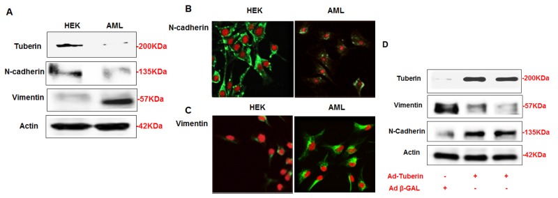
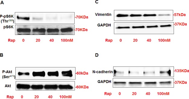
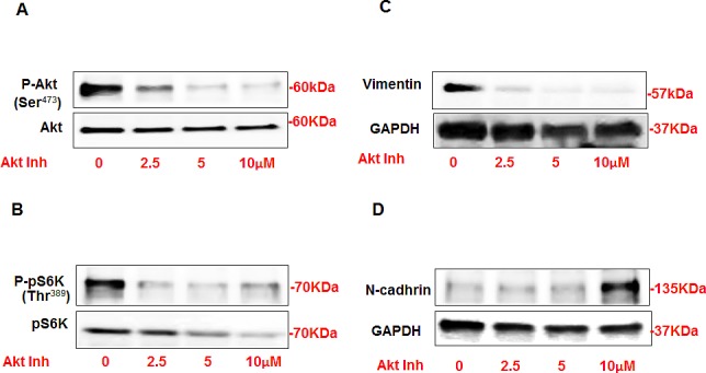
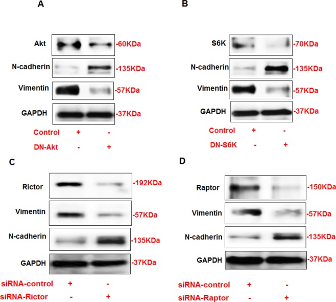
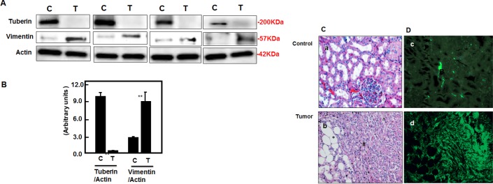
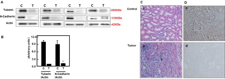
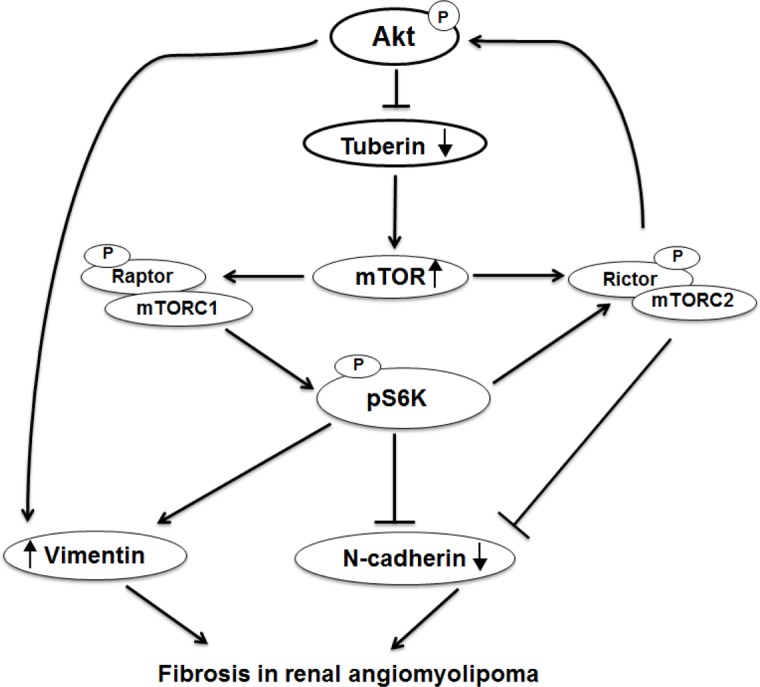
Angiomyolipomas (AMLs) are associated with cell fibrosis in kidney of Tuberous Sclerosis Complex patients. The mechanism by which the fibrotic proteins accumulated in AMLs has not been explored. In the present study, we investigated the role of Akt/tuberin/mTOR pathway in the regulation cell fibrosis proteins. AML cells that expressed low levels of tuberin showed less expression of N-cadherin and higher of vimentin proteins compared to HEK293 cells. AML cells infected with Ad-tuberin showed a significant decrease in vimentin and an increase in N-cadherin protein expression. In addition, cells treated with rapamycin showed a significant increase in p-Akt and a decrease in p-p70S6K that was associated with a decrease expression of vimentin and a slight increase expression in N-cadherin. On the other hand, cells treated with Akt inhibitor revealed a significant decrease in p-Akt and p-p70S6K that was associated with a significant decrease in vimentin and an increase in N-cadherin expression. In addition, cells transfected with DN-Akt or DN-S6K show significant increase expression in N-cadherin and a decrease in vimentin. Moreover, cells transfected with siRNA against rictor or siRNA against raptor resulted in a decrease in vimentin and an increase N-cadherin expression. Kidney tumors from TSC patients showed significant decrease in N-cadherin and significant increased in vimentin protein expression compared to control kidney tissues. These data comprise the first report to provide the role of Akt/tuberin/mTORC1/2 in the regulation of N-cadherin and vimentin that are involved in the progression of fibrosis in kidney tumor of TSC patients.


Conflict of interest statement

Conflict of interest

The authors declare no conflict of interest.

Figures

Figure 1
Figure 1. AML cells express less of N-cadherin and higher of vimentin proteins compared to HEK293 cells
(A) Cells lysates from AML and HEK293 cells were subjected Western blot analysis. Immunoblot analysis show increased in vimentin and decreased in N-cadherin protein expression in AML cells compared to HEK293 cells. (B) AML and HEK293 cells were immunostained for vimentin and N-cadherin using double fluorescence labeling method. The cells were incubated with rabbit antibody against vimentin or N-cadherin followed by secondary anti-rabbit IgG conjugated with FITC. The cells were reacted with Vectashield Mounting Medium with Propedium Iodide (PI) for nuclear staining. (B&C) FITC green signals for N-cadherin and vimentin were detected using a filter with excitation range of 488 nm and PI red signals for nuclear DNA using a filter with excitation at 535 nm. Overlay of vimentin or N-cadherin and DNA staining, demonstrating cell membrane staining for N-cadherin and cytoplasmic staining for vimentin in AML cells. To show staining specificity, control cells were stained without primary antibody. (D) Upregulation of tuberin resulted in decrease in vimentin and increase N-cadherin expression in AML cells. AML cells were infected with adenovirus 6.01 expressing tuberin complementary DNA. An adenovirus vector expressing protein (Adb-GAL) was used as a control. Immunoblot analysis shows overexpression of tuberin decreases vimentin and increases N-cadherin protein expression. Actin was used as a loading control.
Figure 2
Figure 2. Rapamycin treatment significantly decreased P-p70S6K at Thr389 and increased p-Akt at Ser473 that associated with decreased vimentin and increased N-cadherin expression in AML cells
AML cells were treated with different concentrations of rapamycin (0-100nM) for 24h. Western blot analysis was performed in cell lysates using p-p70S6K, p70S6k, p-Akt, Akt, vimentin, N-cadherin and GAPDH antibodies. Cells treated with rapamycin showed significant decrease in protein expression of (A) p-p70S6K and vimentin. On the other hand, significant increase in (B) p-Akt expression and slight increase in (D) N-cadherin showed in cells treated with rapamycin compared to non-treated cells. GAPDH was used as a loading control.
Figure 3
Figure 3. Inhibition of Akt resulted in significant decrease in vimentin and increase in N-cadherin expression in AML cells
AML cells were treated with different concentrations of Akt inhibitor IV (0-10μM) for 24h. Western blot analysis was performed in cell lysates using p-Akt, Akt, p-p70S6K, p70S6k, vimentin, N-cadherin and GAPDH antibodies. Cells treated with Akt inhibitor showed significant decrease in (A) P-Akt at Ser473 and (B) p-p70S6K at Thr389 compared to non-treated cells. Decrease in Akt phosphorylation is associated with significant decrease in (C) vimentin expression in cells treated with lower dose 2.5μM of Akt inhibitor. (D) Cells treated with lower dose of Akt inhibitor (2.5-5 μM) did not show any alteration in N-cadherin while cells treated with higher dose (10 μM) showed significant increase in N-cadherin expression compared to no-treated cells. GAPDH was used as a loading control.
Figure 4
Figure 4. Opposite regulation of N-cadherin and vimentin by DN-Akt and DN-S6K in AML cells
(A) Immunoblot analysis shows that downregulation of Akt by DN-Akt results in an increased in N-cadherin and decreased in vimentin protein expression in AML. In addition, downregulation of S6K by DN-S6K results in decreased in vimentin and increased in N-cadherin protein expression in AML cells. Rictor and Raptor regulates N-cadherin and vimentin (C) Immunoblot analysis shows that downregulation of rictor by siRNA directed against rictor results in decreased in vimentin expression and increased in N-cadherin protein expression in AML cells. In addition, downregulation of raptor by siRNA directed against raptor (D) results in decreased in vimentin expression and increased in N-cadherin protein expression in AML cells. GAPDH was used as a loading control.
Figure 5
Figure 5. Deficiency of tuberin results in increased vimentin protein expression in tumor kidney tissue of TSC patients
(A) Representative Immunoblot analysis showed significant decreased in tuberin and increased in vimentin protein expression in tumor kidney (T) from patients with tuberous sclerosis compared to normal kidney tissues. Actin was used as loading control. (B) Histograms represent means±SE (n=6). Significant difference from control is indicated by **P<0.01. (C) H&E staining shows (a) a normal tubular and glomerular structure in control kidney tissue and (b) *fat, ^vessel and # smooth muscle cells types in kidney angiomyolipoma tissue of TSC patients. (D) Kidney sections were stained with vimentin followed by immunofluroscene staining. Control of kidney (c) shows a few cells stained with vimentin while (d) most of blood vessel and smooth muscle cells were stained in kidney tumor tissue. Control sections in both procedures were incubated without primary antibody.
Figure 6
Figure 6. Deficiency of tuberin results in decreased N-cadherin protein expression in tumor kidney tissue of TSC patients
(A) Representative Immunoblot analysis showed significant decreased in tuberin and decreased in N-cadherin protein expression in tumor kidney (T) from patients with tuberous sclerosis compared to normal kidney tissues. Actin was used as loading control. (B) Histograms represent means±SE (n=6). Significant difference from control is indicated by **P<0.01. (C) H&E staining shows (a) a normal tubular and glomerular structure in control kidney tissue and (b) *fat, ^vessel and # smooth muscle cells types in kidney angiomyolipoma tissue of TSC patients. (D) Kidney sections were stained with N-cadherin followed by horseradish peroxidase staining. Control of kidney (c) shows a few cells stained with N-cadherin while (d) most of blood vessel and smooth muscle cells were stained in kidney tumor tissue. Control sections in both procedures were incubated without primary antibody.
Figure 7
Figure 7. Proposed model of regulation of N-cadherin and vimentin in kidney angiomyolipoma
Deficiency in tuberin upregulates vimentin and downregulates N-cadherin in kidney tumor of TSC patients. In addition, activation of Akt enhances vimnetin and decreases N-cadherin expression while activation of mTORC1/2 increases vimentin and decreases N-cadherin resulted in cell fibrosis progression in renal angiomyolipoma of tuberous sclerosis patients.

References

    1. Bjornsson J, Short MP, Kwiatkowski DJ, Henske EP. Tuberous sclerosis-associated renal cell carcinoma: clinical, pathological, and genetic features. Am J Pathol. 1996;149:1201–1208. - PMC - PubMed
    1. Al-Saleem T, Wessner LL, Scheithauer BW, Patterson K, Roach ES, Dreyer SJ, Fujikawa K, Bjornsson J, Bernstein J, Henske EP. Malignant tumors of the kidney, brain, and soft tissues in children and young adults with the tuberous sclerosis complex. Cancer. 1998;83:2208–2216. - PubMed
    1. Parry L, Maynard JH, Patel A, Clifford SC, Morrissey C, Maher ER, Cheadle JP, Sampson JR. Analysis of the TSC1 and TSC2 genes in sporadic renal cell carcinomas. Br J Cancer. 2001;85:1226–1230. - PMC - PubMed
    1. Henske EP, Neumann HP, Scheithauer BW, Herbst EW, Short MP, et al. Loss of heterozygosity in the tuberous sclerosis (TSC2) region of chromosome band 16p13 occurs in sporadic as well as TSC-associated renal angiomyolipomas. Genes Chromosomes Canc. 1995;13:295–298. - PubMed
    1. Rennebeck G, Kleymenova EV, Anderson R, Yeung RS, Artzt K, Walker CL. Loss of function of the tuberous sclerosis 2 tumor suppressor gene results in embryoni c lethality characterized by disrupted neuroepithelial growth and development. Proc Natl Acad Sci USA. 1998;95:15629–15634. - PMC - PubMed

Publication types

MeSH terms