. 2014 Oct 15;307(8):L618-31.
doi: 10.1152/ajplung.00144.2014. Epub 2014 Aug 22.

Regulation of fibroblast lipid storage and myofibroblast phenotypes during alveolar septation in mice

Affiliations

Regulation of fibroblast lipid storage and myofibroblast phenotypes during alveolar septation in mice

Stephen E McGowan et al. Am J Physiol Lung Cell Mol Physiol. .

Abstract

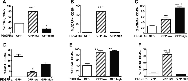
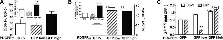
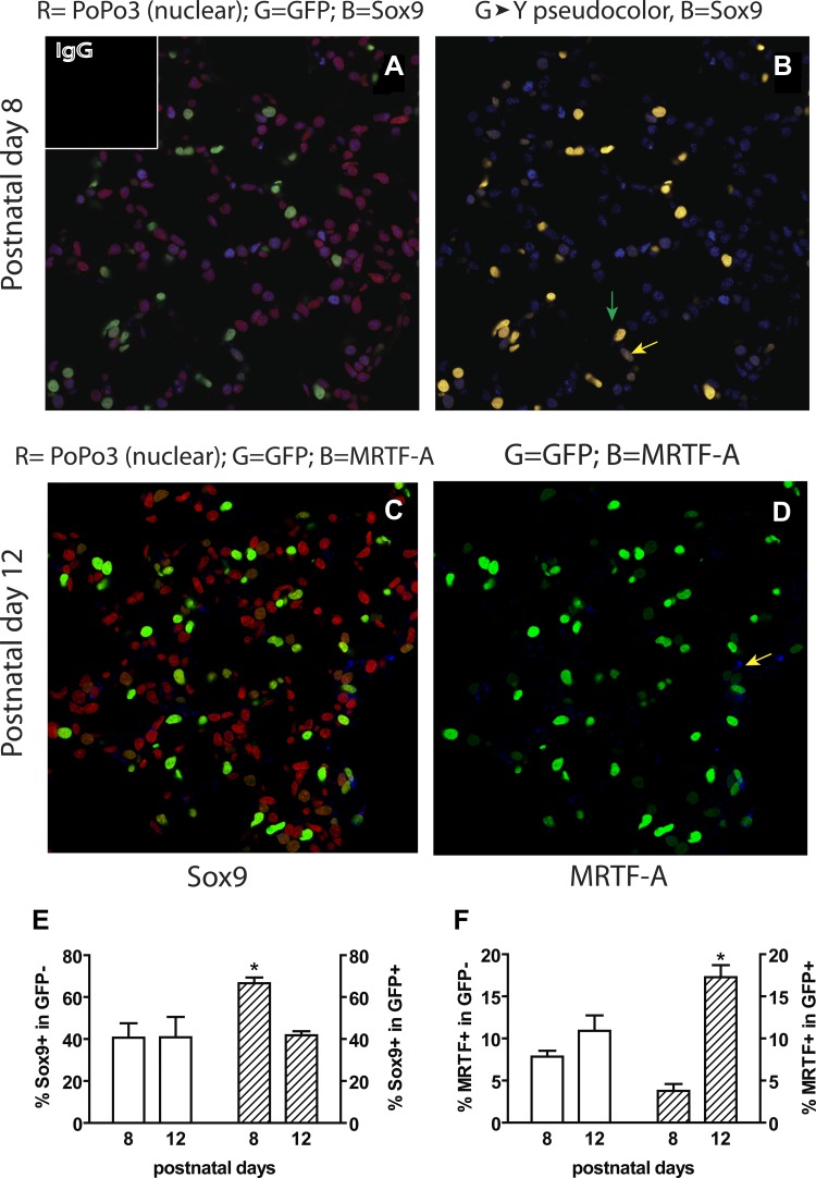
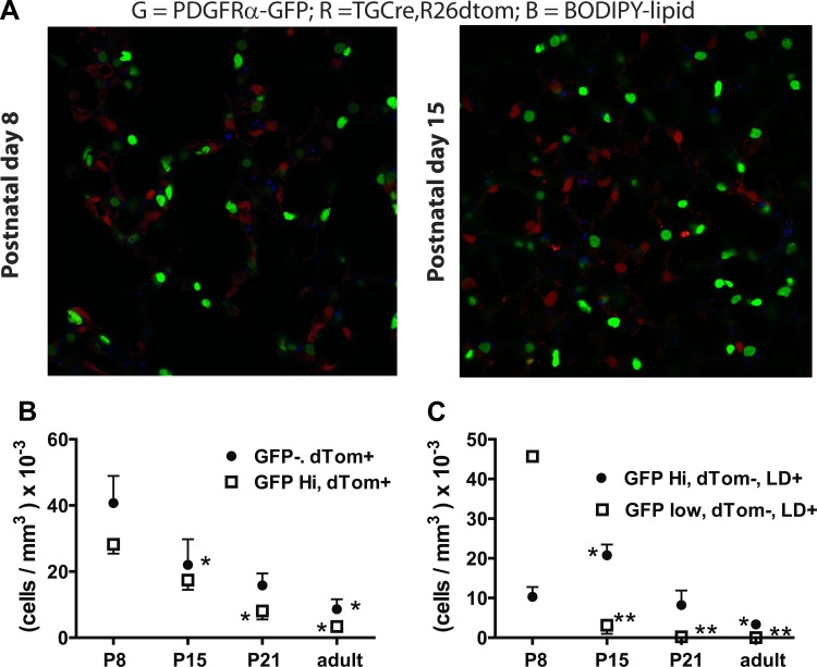
Signaling through platelet-derived growth factor receptor-α (PDGFRα) is required for alveolar septation and participates in alveolar regeneration after pneumonectomy. In both adipose tissue and skeletal muscle, bipotent pdgfrα-expressing progenitors expressing delta-like ligand-1 or sex-determining region Y box 9 (Sox9) may differentiate into either lipid storage cells or myofibroblasts. We analyzed markers of mesenchymal progenitors and differentiation in lung fibroblasts (LF) with different levels (absent, low, or high) of pdgfrα gene expression. A larger proportion of pdgfrα-expressing than nonexpressing LF contained Sox9. Neutral lipids, CD166, and Tcf21 were more abundant in LF with a lower compared with a higher level of pdgfrα gene expression. PDGF-A increased Sox9 in primary LF cultures, suggesting that active signaling through PDGFRα is required to maintain Sox9. As alveolar septation progresses from postnatal day (P) 8 to P12, fewer pdgfrα-expressing LF contain Sox9, whereas more of these LF contain myocardin-like transcription factor-A, showing that Sox9 diminishes as LF become myofibroblasts. At P8, neutral lipid droplets predominate in LF with the lower level of pdgfrα gene expression, whereas transgelin (tagln) was predominantly expressed in LF with higher pdgfrα gene expression. Targeted deletion of pdgfrα in LF, which expressed tagln, reduced Sox9 in α-actin (α-SMA, ACTA2)-containing LF, whereas it increased the abundance of cell surface delta-like protein-1 (as well as peroxisome proliferator-activated receptor-γ and tcf21 mRNA in LF, which also expressed stem cell antigen-1). Thus pdgfrα deletion differentially alters delta-like protein-1 and Sox9, suggesting that targeting different downstream pathways in PDGF-A-responsive LF could identify strategies that promote lung regeneration without initiating fibrosis.

Keywords: adipocyte; delta-like ligand-1 (Dlk1); lung alveolarization; platelet-derived growth factor; preadipocyte factor-1; sex-determining region Y box 9; stem cell; stereology.


Figures

Fig. 1.
Fig. 1.
Genetic manipulations used to label alveolar mesenchymal cells. Platelet-derived growth factor receptor (PDGFR) α-expressing lung fibroblasts (LF) were marked with enhanced green fluorescent protein (GFP), regulated by the endogenous PDGFRα promoter (PDGFRα-GFP, A). Cell surface CD140a (PDGFRα) increased with GFP fluorescence, and PDGFREα mRNA was more abundant in the GFPhigh cells. **P < 0.01, GFPlow or GFPhigh compared with GFP negative (GFP). *P < 0.05, GFPhigh compared with GFPlow. †P < 0.05, GFPhigh vs. GFPlow. B: 2 subpopulations were distinguished based on the intensity of GFP fluorescence: GFPhigh LF, which reside more distally in the elongating alveolar septa, and GFPlow LF, which contain lipid droplets that reside at the base of septa at postnatal day (P) 8 but regress by P15. C: a separate line of mice that expressed Cre recombinase controlled by the transgelin (TG) promoter was used to excise a stop codon and enable expression of tdTomato (dTom), which was expressed in pericytes and at P8 and in PDGFRα-GFP LF: TGCre;R26Tom; PDGFRα-GFP (contain both GFP and dTom, B). TG-Cre was also used to inactivate LoxP-flanked PDGFRα in cells where TG expression is abundant, which has functional consequences in PDGFRα-expressing LF but not in pericytes, which express PDGFRβ but not PDGFRα.
Fig. 2.
Fig. 2.
Representative plots from flow cytometric (FACS) analysis of LF. Pdgfrα-expressing (GFP+) and -nonexpressing (GFP) LF were isolated from mice bearing the pdgfrα-driven GFP marker. A: representative dot plots showing the distribution of events for LF that are GFPlow, GFPhigh, or are not expressing PDGFRα GFP. Panels a, e, g, i, and k show staining with isotype control immunoglobulins (IgG), whereas the remaining panels show staining with the designated immune IgGs or LipidTOX red (LTR). All adherent cells meeting the gating criteria for side scatter and forward scatter width are shown. CD45+ cells were not excluded from these plots but were excluded for the combined data shown in the bar graphs in Figs. 3–6. The numbers show percentage of total gated cells (R2 in Bb) in the three populations distinguished by GFP intensity. B: gating strategy used to evaluate Sca1 and CD34 in CD45 LF. Forward scatter area (FSC-A) vs. side scatter area (SSC-A) (a) or forward scatter width (FSC-W) (b) were used to eliminate potential aggregates or small dead cells. c, Negative selection using allophycocyanin (APC)-anti-CD45 identified LF with different levels of GFP fluorescence (R3, R4, R5). CD45+ LF were only observed in the GFP population and were excluded from subsequent analyses. df, LF stained with Brilliant violet (Brill-Viol 421) anti-CD34 and APC-Cy7 anti-Sca1 within the 3 CD45 populations (R3-R5). Percentages of total CD45 cells in each quadrant are shown.
Fig. 3.
Fig. 3.
FACS analysis of progenitor markers in fibroblasts (LF) with different levels of pdgfrα gene expression. LF isolated from PDGFRα-GFP mice at P8 were stained for the antigens shown on the y-axes: LTR, neutral lipid binding LTR (A); ADRP, adipocyte differentiation related protein, perilipin-2 (B); α-SMA, α-smooth muscle actin, acta2; (C); Sca1, stem cell antigen-1 (D); CD34, podocalyxin (E); and CD166, activated leukocyte adhesion molecule (ALCAM), activated-leukocyte cell adhesion molecule (F); the CD45 cell populations were selected as shown in Fig. 2 and expressed as a percentage of all CD45 LF within a particular population based on the intensity of GFP fluorescence (GFP, GFPlow, GFPhigh). **P < 0.01 and *P < 0.05, mean ± SE, n = 6 mice, 2-way ANOVA, Student-Newman-Keuls post hoc test comparing GFPlow or GFPhigh LF with GFP. †P < 0.01 comparing GFPlow with GFPhigh.
Fig. 4.
Fig. 4.
Ki-67 is diminished, whereas p57kip2, G0S2, and Tcf21 are increased in GFPlow LF. FACS analysis of intracellular proteins [A, Ki67 (MKI67) P4; B, Ki67 P8; C, p57kip2 cyclin-dependent kinase inhibitor (CDKN1c); D, G0-G1 switch protein-2 (G0S2); E, Tcf21 (Pod1)] were analyzed by FACS at P8, except for A which shows LF at P4. The proportions of CD45 LF containing the various antigens are expressed as a percentage of all CD45 within a particular population. F: quantitative real-time PCR (qRT-PCR, normalizing to constitutive β2-microglobulin expression) analysis of G0S2 and Tcf21 mRNA in GFPlow and GFPhigh expressed relative to PDGFRα-GFP LF obtained after sorting by FACS (n = 4). **P < 0.01 and *P < 0.05, mean ± SE, n = 4 mice, 2-way ANOVA, Student-Newman-Keuls post hoc test comparing GFPlow or GFPhigh LF with GFP. †P < 0.05 and ††P < 0.01, comparing GFPlow with GFPhigh.
Fig. 5.
Fig. 5.
The abundance of sex-determining region Y box 9 (Sox9) correlates with pdgfrα gene expression. FACS analysis of the 3 LF subpopulations bearing cell surface delta-like protein-1 (Dlk1) (n = 7) (A) and intracellular Sox9 (n = 5) (B) were assessed at P8 using FACS. The proportions of CD45 LF containing Dlk1 or Sox9 are expressed as a percentage of all CD45 within a particular population. Insets show the cells that are both Sca1+ and CD45 relative to the total CD45 LF within each subpopulation defined by GFP intensity. C: qRT-PCR showing Sox9 and Dlk1 mRNA in GFPlow and GFPhigh expressed relative to PDGFRα-GFP LF (n = 4). **P < 0.01 and *P < 0.05, mean ± SE, 2-way ANOVA, Student-Newman-Keuls post hoc test comparing GFPlow or GFPhigh LF with GFP. †P < 0.01, comparing GFPlow with GFPhigh.
Fig. 6.
Fig. 6.
Targeted pdgfrα gene deletion alters the abundance of Dlk1, Sox9, and Tcf21. AD: FACS analysis of LF isolated at P8 from 5 mice bearing the transgelin-mediated (TG-Cre) conditional deletion of loxP-flanked pdgfrα (PDGFRαF/F) or 5 littermate controls, gating on CD45 cells. A: %Dlk1+, LTR+, or Dlk1+,Sca1+ gating on CD45 (left); or %LTR+ within CD45 (axis on right). B: Dlk1+ LF within the CD45, CD34+ population (axis on left); %CD45, CD34+ relative to all CD45 LF (axis on right). C: %CD166+ (axis on left) or both CD166+, Dlk1+ (axis on right) in the CD45 population. *P < 0.05, n = 5, mean ± SE, t-test, paired variables. D: %CD45 LF containing α-SMA in control (open bars) or TGCre+/−;PDGFRαF/F (solid bars, axis on right). %Sox9+, CD45, α-SMA+ subpopulation, expressed relative to all α-SMA+ LF (axis on left). E: qRT-PCR analysis of periostin (Postn), periostin-lacking exon 21 (Postn 20–21), tcf21, and pparγ in PDGFRα-deleted and controls within the same litter, n = 5 litters. *P < 0.05 and **P < 0.01, mean ± SE, t-test, paired variables comparing PDGFRαF/F LF with those from their respective littermate controls. F: qRT-PCR using a probe that is not influenced by splicing dlk1-T (total), the A and C splice variants (dlk1-A and dlk1-C, respectively) comparing control and TGCre+/−;PDGFRαF/F mice (mean ± SE, n = 3 for each group); *P < 0.05, n = 3. dlk1-T was normalized to β2-microglobulin mRNA, whereas the dlk1-A and -C splice variants were normalized to dlk1-T for each respective sample.
Fig. 7.
Fig. 7.
PDGFR-A increases Sox9 mRNA and protein in cultured LF. Primary isolates of LF were cultured and exposed to 20 ng PDGF-AA or 2.5 ng TGF-β1/ml for 24 h. A: RNA was isolated, and quantitative real-time PCR was performed using TaqMan probes. mRNA was compared with LF cultured in the absence of PDGF or TGF-β1 (control) within each respective experiment using the 2−(ΔΔCT) method. Representative immunoblots are shown after probing for Sox9 (B) and myocardin-like transcription factor-A (MRTF-A, C) using β-tubulin (βTub) as a loading control. The densities for Sox9 and MRTF-A were compared relative to the density of the corresponding unexposed control culture after normalizing for the density of βtub for each respective condition; n = 6 independent experiments using different primary LF isolations for both RNA and protein. *P < 0.05, mean ± SE, 1-way ANOVA, Student-Newman-Keuls post hoc test.
Fig. 8.
Fig. 8.
Sox9 decreases, whereas MRTF-A increases, during septal elongation. Lungs from PDGFRα-GFP+ mice were uniformly inflated and fixed at P8 or P12 and immunostained for Sox9 or MRTF-A; n = 4 or 3 mice, respectively. A and B: nuclei containing Sox9 (blue), which is more readily observed in B, where green was changed to a yellow pseudocolor for better contrast. Yellow and green arrows show GFPlow and GFPhigh nuclei, respectively, containing Sox9. C and D: MRTF-A (blue) surrounding nuclei (GFP+, arrow). The proportions of pdgfrα-expressing (GFP+) or -nonexpressing (GFP) alveolar cells within their respective parent populations (all GFP+ LF, hatched bars, axes on right; or GFP LF, open bars, axes on left) at P8 were compared with those at P12 for Sox9 or MRTF-A in E or F, respectively. *P < 0.05, mean ± SE, 3-way ANOVA, Student-Newman-Keuls post hoc test.
Fig. 9.
Fig. 9.
Lipid droplets and transgelin (TG) are observed in different LF subpopulations. Lungs from four TG Cre+/−;Rosa26dTomato (Tom)+/−;PDGFRαGFP+ mice at various postnatal days or adults were stained with BODIPY 665/670 (to visualize neutral lipids) and imaged using confocal microscopy (A). Alveolar cells containing dTomato (dTom+) and/or GFP were enumerated and expressed/mm3 of lung tissue. B: only GFP-bright (high, Hi) cells are shown because no GFP-dim (low) cells also contained dTom (expressed transgelin). C: only cells not expressing transgelin (dTom) contained BODIPY 665/676-staining lipid droplets (LD). LD+ cells were distinguished and enumerated based on the intensity of GFP fluorescence (bright, Hi or dim, low). *P < 0.05, mean ± SE, n = 4 at each age (**P < 0.01), 1-way ANOVA for age, Student-Newman-Keuls post hoc test comparing P8 with older mice within each determination (shown by same symbol).
Fig. 10.
Fig. 10.
pdgfrα gene deletion alters respiratory mechanics. Representative microscopic fields for control (A) and TGCre+/−;PDGFRαF/F (B) mice; magnification ×50. Insets, elastic fibers in the alveolar walls (arrows), which were thinner and disconnected in mice with PDGFRα disruption. C: static pressure-volume curves for control (n = 8, closed circles) and TGCre+/−;PDGFRαF/F (n = 5, open squares) mice showing that targeted pdgfrα gene deletion increased the compliance of the respiratory system (combined lung and chest wall).

References

    1. Acharya A, Baek ST, Huang G, Eskiocak B, Goetsch S, Sung CY, Banfi S, Sauer MF, Olsen GS, Duffield JS, Olson EN, Tallquist MD. The bHLH transcription factor Tcf21 is required for lineage-specific EMT of cardiac fibroblast progenitors. Development 139: 2139–2149, 2012 - PMC - PubMed
    1. Andersen DC, Laborda J, Baladron V, Kassem M, Sheikh SP, Jensen CH. Dual role of delta-like 1 homolog (DLK1) in skeletal muscle development and adult muscle regeneration. Development 140: 3743–3753, 2013 - PubMed
    1. Berry R, Rodeheffer MS. Characterization of the adipocyte cellular lineage in vivo. Nat Cell Biol 15: 302–308, 2013 - PMC - PubMed
    1. Boström H, Gritli-Linde A, Betsholtz C. PDGF-A/PDGF alpha-receptor signaling is required for lung growth and the formation of alveoli but not for early lung branching morphogenesis. Dev Dynam 223: 155–162, 2002 - PubMed
    1. Boström H, Willetts K, Pekny M, Leveen P, Lindahl P, Hedstrand H, Pekna M, Hellstrom M, Gebre-Medin S, Schalling M, Nilsson M, Kurland S, Tornell J, Heath JK, Betsholtz C. PGDF-A signaling is a critical event in lung alveolar myofibroblast development and alveogenesis. Cell 85: 863–873, 1996 - PubMed

Publication types

MeSH terms

LinkOut - more resources