. 2014 Nov 1;127(Pt 21):4714-27.
doi: 10.1242/jcs.155275. Epub 2014 Aug 22.

NPHP4 controls ciliary trafficking of membrane proteins and large soluble proteins at the transition zone

Affiliations

NPHP4 controls ciliary trafficking of membrane proteins and large soluble proteins at the transition zone

Junya Awata et al. J Cell Sci. .

Abstract

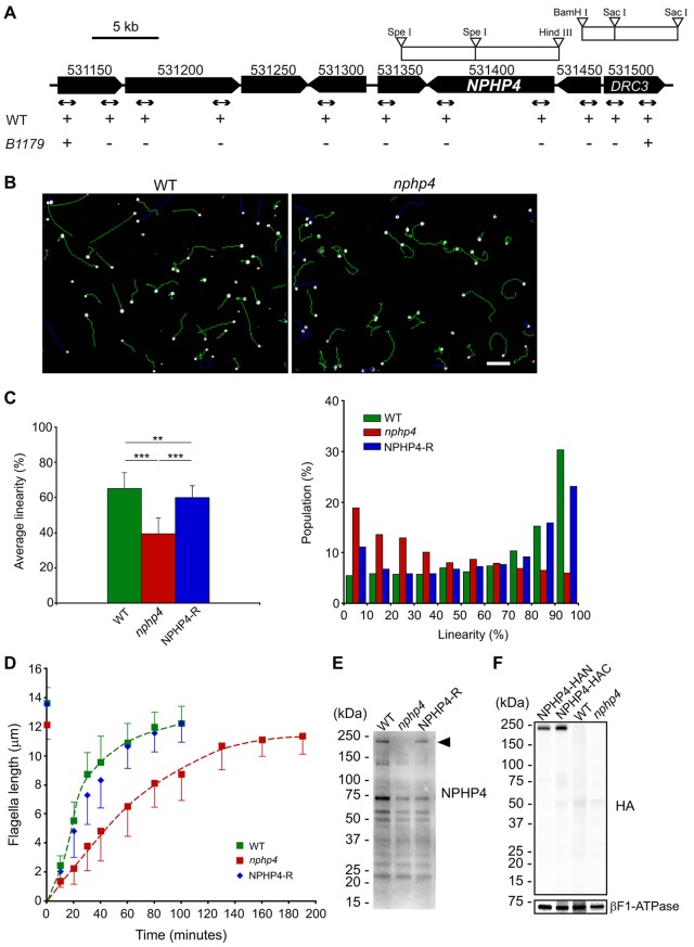
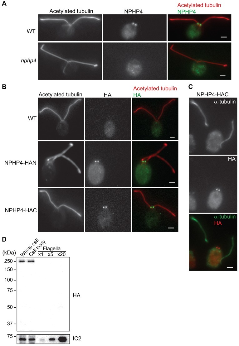
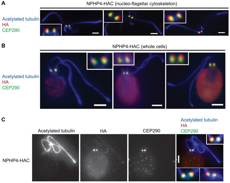
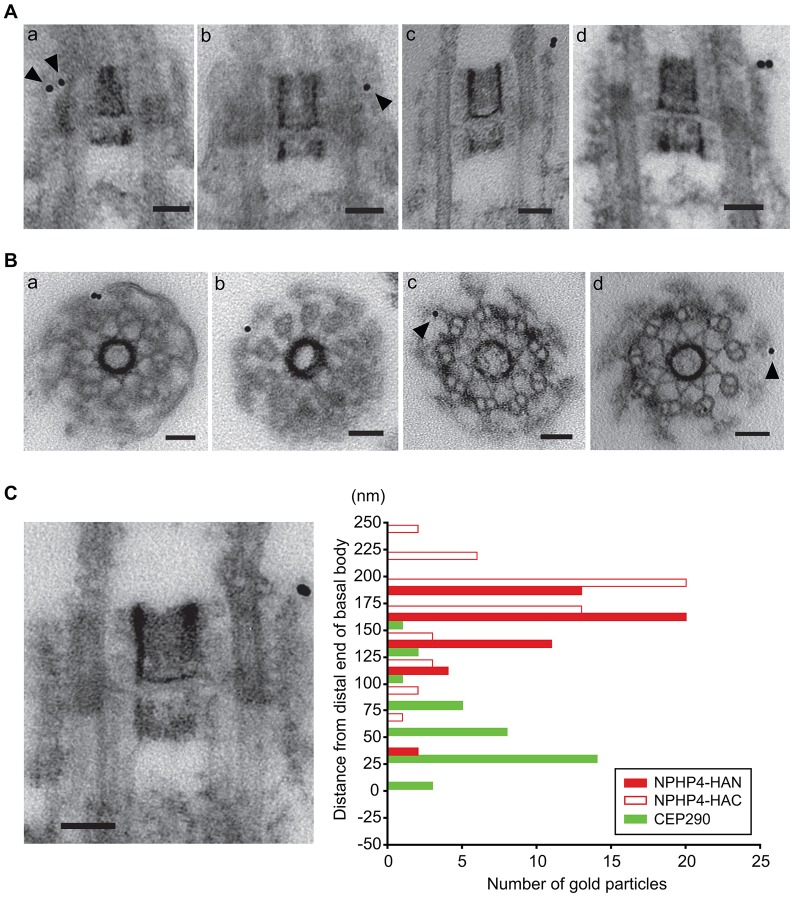
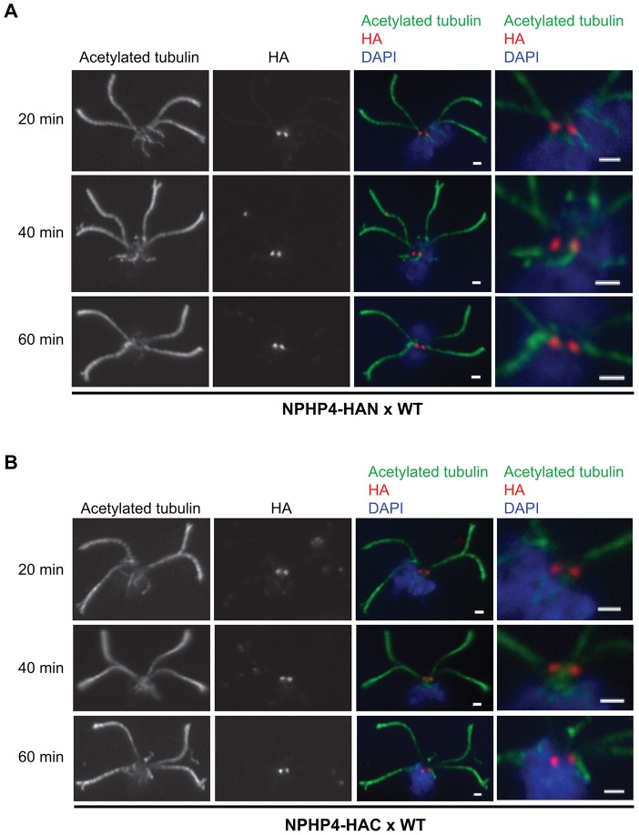
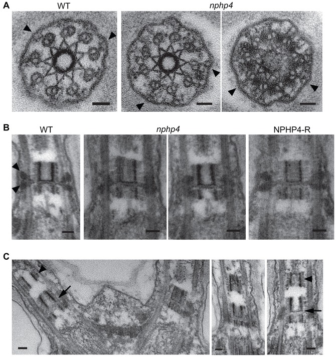
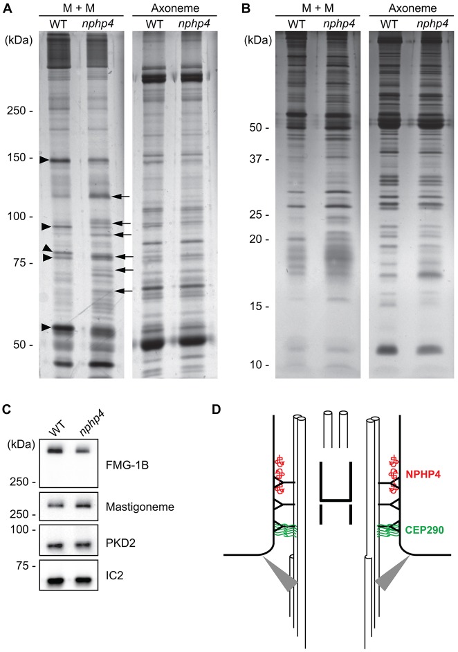
The protein nephrocystin-4 (NPHP4) is widespread in ciliated organisms, and defects in NPHP4 cause nephronophthisis and blindness in humans. To learn more about the function of NPHP4, we have studied it in Chlamydomonas reinhardtii. NPHP4 is stably incorporated into the distal part of the flagellar transition zone, close to the membrane and distal to CEP290, another transition zone protein. Therefore, these two proteins, which are incorporated into the transition zone independently of each other, define different domains of the transition zone. An nphp4-null mutant forms flagella with nearly normal length, ultrastructure and intraflagellar transport. When fractions from isolated wild-type and nphp4 flagella were compared, few differences were observed between the axonemes, but the amounts of certain membrane proteins were greatly reduced in the mutant flagella, and cellular housekeeping proteins >50 kDa were no longer excluded from mutant flagella. Therefore, NPHP4 functions at the transition zone as an essential part of a barrier that regulates both membrane and soluble protein composition of flagella. The phenotypic consequences of NPHP4 mutations in humans likely follow from protein mislocalization due to defects in the transition zone barrier.

Keywords: CEP290; Chlamydomonas; Cilia; Flagella; Nephrocystin-4; Transition zone.


Figures

Fig. 1.
Fig. 1.
Characterization of the C. reinhardtii nphp4 mutant. (A) Map of C. reinhardtii genome near the NPHP4 locus. Numbers above each locus correspond to gene IDs in Phytozome version 9.1 (http://www.phytozome.net). Arrows indicate the positions of PCR products used to delimit the deleted region; plus and minus marks indicate whether the PCR products were amplified. Genome fragments from bacterial artificial chromosome (BAC) clones used for knock-in of full-length NPHP4 and DRC3 are indicated by white rectangles. WT, wild type. (B) Representative images showing swimming paths of wild-type and nphp4 cells. White dots show cells where CASA began to monitor their tracks; green lines are the swimming paths analyzed. Blue tracks were not analyzed because the cells swam outside the microscope field before recording was completed. Cells marked with red dots were immobilized by attachment to the coverslip. The swimming paths of nphp4 cells are more erratic than those of wild-type cells. Scale bar: 50 µm. (C) Bar graph (left) showing the mean±s.d. of linearity </emph>and histogram (right) summarizing the distribution of linearity in swimming paths of populations of wild-type, nphp4 and NPHP4-R cells (see supplementary material Fig. S1). For each strain, the population was calculated from the sum of values from a total of ten fields in each of five independent experiments. Statistical significance was determined by the Tukey–Kramer method: **P<0.01; ***P<0.001. (D) Flagella length prior to deflagellation and as a function of time after deflagellation of wild-type, nphp4 and NPHP4-R cells. Flagellar length at each time-point is the mean±s.d. of 30 flagella. (E) Western blot of whole cells probed with an antibody against the C-terminus of NPHP4. The antibody recognized a band (arrowhead) of the predicted size for NPHP4 (210 kDa) in wild-type cells and the NPHP4-R strain. (F) Western blot of wild-type, nphp4, NPHP4–HAN and NPHP4–HAC whole cells probed with a monoclonal antibody against the HA peptide. βF1-ATPase was used as a loading control.
Fig. 2.
Fig. 2.
NPHP4 is localized to the base of flagella. (A) Wild-type (WT) and nphp4 cells were labeled with antibodies against acetylated tubulin (a marker for flagellar microtubules) and NPHP4; merged images are on the right. (B) Wild-type, NPHP4–HAN and NPHP4–HAC cells were labeled with anti-acetylated tubulin and anti-HA; merged images are on the right. (C) Detached flagella and cell body of an NPHP4–HAC cell fixed and labeled with antibodies against α-tubulin and the HA peptide. Scale bars: 2 µm. (D) NPHP4–HAC whole cells, cell bodies and isolated flagella were analyzed on a western blot probed with the anti-HA antibody; the axonemal protein IC2 was used as a loading control. ×1, ×5 and ×20 indicate that ∼1, 5 or 20 flagella pairs were loaded per cell body.
Fig. 3.
Fig. 3.
NPHP4 is localized to the transition zone. Isolated nucleoflagellar apparatuses of NPHP4–HAC cells (A) or whole NPHP4–HAC cells (B,C) were labeled with antibodies against acetylated tubulin, HA peptide and CEP290. Images were acquired by conventional widefield immunofluorescence microscopy (A,B) or TESM (C). All the images in A and B and the image in the right panel of C are merged from separate channels (shown only in C). The insets in A and B and the upper inset in C are enlargements of the transition zones shown in the panels; the lower insets in C show two additional examples. Scale bars: 2 µm.
Fig. 4.
Fig. 4.
NPHP4 is located in the distal transition zone. Isolated nucleoflagellar apparatuses of NPHP4–HAN or NPHP4–HAC cells were incubated with rat IgG directed against the HA peptide, followed by incubation with anti-rat-IgG conjugated to 10-nm gold particles. (A) Typical longitudinal sections of NPHP4–HAN (a,b) and NPHP4–HAC (c,d) transition zones labeled with gold particles (arrowheads). (B) Cross-sections of NPHP4–HAN (a,b) and NPHP–HAC (c,d) transition zones labeled with gold particles (arrowheads). All gold particles were found at the periphery of the transition zone, in close association with the transition zone membrane. (C) Distribution of gold particles associated with longitudinal sections of NPHP4–HAN and NPHP4–HAC transition zones; 50 gold particles were scored for each. The electron micrograph shows a typical example matched to the scale of the y-axis; ‘0’ on the y-axis indicates the boundary (visible in the longitudinal section) between the basal body and the transition zone. The C-terminus of NPHP4 might be located slightly more distally than the N-terminus. The green bars show the distribution of CEP290 replotted from Craige et al. (Craige et al., 2010). Scale bars: 50 nm.
Fig. 5.
Fig. 5.
NPHP4 at the transition zone is static. Wild-type (WT) gametes were mixed with NPHP4–HAN (A) or NPHP4–HAC (B) gametes to initiate the mating reaction. At the indicated times after mixing, nucleoflagellar apparatuses of the resulting quadriflagellated zygotes were prepared, fixed and stained with anti-acetylated tubulin, anti-HA and DAPI. The right panels are enlargements of the transition zones shown in the merged images. Scale bars: 1 µm.
Fig. 6.
Fig. 6.
Transition zone structure in the nphp4 mutant. (A) Cross-sections through transition zones of wild-type (WT) and nphp4 cells. The Y-links (arrowheads) appear normal in the nphp4 mutant. (B) Longitudinal sections through transition zones of wild-type, nphp4 and NPHP4-R cells. The electron-opaque wedge-shaped structures (arrowheads) are present in wild-type and NPHP4-R transition zones, but are missing, deformed or obscured in the nphp4 transition zones. (C) About one-third of nphp4 flagella contain ectopic cylindrical structures (arrowheads) just distal to the transition zone; these structures resemble the normal transition zone cylinders (arrows). Scale bars: 50 nm.
Fig. 7.
Fig. 7.
Flagella of nphp4 cells have abnormal protein composition. 6% (A) or 12% (B) SDS-polyacrylamide gels were used to compare wild-type (WT) versus nphp4 membrane-plus-matrix (M+M) and axonemal fractions. Arrowheads indicate some proteins that decreased in amount in the nphp4 membrane-plus-matrix fraction; arrows indicate some proteins that increased in amount in the mutant flagella. (C) Western blot of isolated wild-type and nphp4 flagella probed with antibodies against FMG-1B, the mastigoneme protein and PKD2. The outer arm dynein intermediate chain IC2 served as a loading control. (D) Diagram illustrating the distribution of NPHP4 and CEP290 in the transition zone. NPHP4 (red) is located in close association with the membrane at the distal end of the transition zone. CEP290 (green) is located in the space between the doublet microtubules and membrane at the proximal end of the transition zone. Gray triangles represent transition fibers connecting the basal body to the plasma membrane.

Similar articles

Cited by

References

    1. Atteia A., van Lis R., Gelius-Dietrich G., Adrait A., Garin J., Joyard J., Rolland N., Martin W. (2006). Pyruvate formate-lyase and a novel route of eukaryotic ATP synthesis in Chlamydomonas mitochondria. J. Biol. Chem. 281, 9909–9918. 10.1074/jbc.M507862200 - DOI - PubMed
    1. Berthold P., Schmitt R., Mages W. (2002). An engineered Streptomyces hygroscopicus aph 7″ gene mediates dominant resistance against hygromycin B in Chlamydomonas reinhardtii. Protist 153, 401–412. 10.1078/14344610260450136 - DOI - PubMed
    1. Bessen M., Fay R. B., Witman G. B. (1980). Calcium control of waveform in isolated flagellar axonemes of Chlamydomonas. J. Cell Biol. 86, 446–455. 10.1083/jcb.86.2.446 - DOI - PMC - PubMed
    1. Bloodgood R. A., Woodward M. P., Salomonsky N. L. (1986). Redistribution and shedding of flagellar membrane glycoproteins visualized using an anti-carbohydrate monoclonal antibody and concanavalin A. J. Cell Biol. 102, 1797–1812. 10.1083/jcb.102.5.1797 - DOI - PMC - PubMed
    1. Breslow D. K., Koslover E. F., Seydel F., Spakowitz A. J., Nachury M. V. (2013). An in vitro assay for entry into cilia reveals unique properties of the soluble diffusion barrier. J. Cell Biol. 203, 129–147. 10.1083/jcb.201212024 - DOI - PMC - PubMed

Publication types

MeSH terms

Substances

LinkOut - more resources