Review
. 2014 Dec;24(12):734-42.
doi: 10.1016/j.tcb.2014.07.008. Epub 2014 Aug 20.

Damage control: cellular mechanisms of plasma membrane repair

Affiliations
Review

Damage control: cellular mechanisms of plasma membrane repair

Norma W Andrews et al. Trends Cell Biol. 2014 Dec.

Abstract

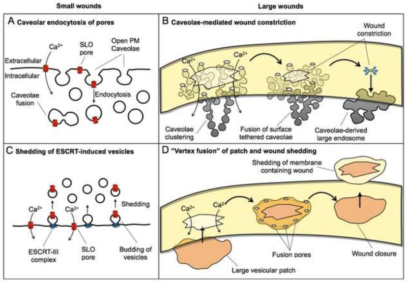
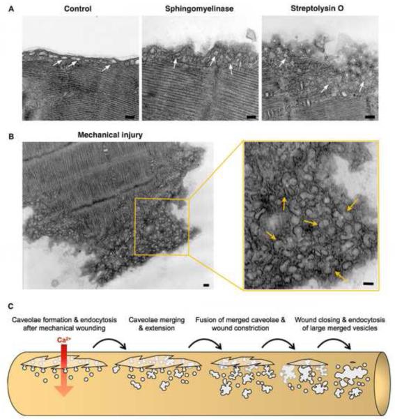
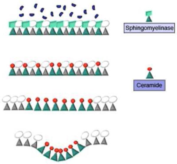
When wounded, eukaryotic cells reseal in a few seconds. Ca(2+) influx induces exocytosis of lysosomes, a process previously thought to promote repair by 'patching' wounds. New evidence suggests that resealing involves direct wound removal. Exocytosis of lysosomal acid sphingomyelinase (ASM) triggers endocytosis of lesions followed by intracellular degradation. Characterization of injury-induced endosomes revealed a role for caveolae, sphingolipid-enriched plasma membrane invaginations that internalize toxin pores and are abundant in mechanically stressed cells. These findings provide a novel mechanistic explanation for the muscle pathology associated with mutations in caveolar proteins. Membrane remodeling by the ESCRT complex was also recently shown to participate in small-wound repair, emphasizing that cell resealing involves previously unrecognized mechanisms for lesion removal that are distinct from the patch model.

Keywords: caveolae; endocytosis; injury; muscular dystrophy; resealing.


Figures

Figure 1
Figure 1. Proposed mechanisms for plasma membrane repair
Small wounds (<100 nm) such as those generated by pore-forming toxins were proposed to be repaired by two mechanisms: (A) endocytosis of caveolar vesicles , and (C) ESCRT-mediated vesicle budding . Large wounds (>100 nm) were proposed to be repaired by: (B) a mechanism involving endocytosis, clustering and fusion of caveolae that leads to wound constriction , and (D) patching by a large intracellular exocytic vesicle, proposed to fuse at several points at the periphery of the wound, resulting in shedding of the damaged membrane . Internalization of SLO pores in caveolar vesicles has been observed , but direct evidence for extracellular shedding of wounded plasma membrane is still not available.
Figure 2
Figure 2. Transmembrane pores formed by SLO are internalized in caveolar vesicles which gradually traffic into lysosomes for degradation
During the first 30 seconds after cell permeabilization with SLO in the presence of Ca2+, individual caveolae accumulate at the cell periphery. In subsequent minutes, caveolar vesicles carrying SLO pores merge, forming larger compartments. SLO pores are ultimately sorted into luminal vesicles of multivesicular bodies, and degraded in mature lysosomes . The images show transmission EM of endocytic vesicles observed in NRK cells at increasing time points after permeabilization with SLO; the arrows point to BSA-gold sued as an endocytic tracer.
Figure 3
Figure 3. Caveolae-like vesicles accumulate below the sarcolemma of primary muscle fibers in response to injury or exposure to purified sphingomyelinase
(A) Transmission EM images of mouse Flexor digitorum brevis muscle fibers untreated (Control), exposed to 50 mU/ml B. cereus sphingomyelinase for 5 min, or treated with 400 ng/ml SLO for 30 seconds. (B) Transmission EM image of the mechanically severed tip of a Flexor digitorum brevis muscle fiber. Arrows in the enlarged section to the right point to merged caveolae-derived compartments below the sarcolemma. Bars= 100 nm. (C) Proposed caveolae-mediated mechanism for the resealing of mechanical wounds on a muscle fiber. Ca2+ flowing through a wound would induce internalization and intracellular merging of sarcolemma-associated caveolae, leading to the formation of larger compartments tethered to the sarcolemma that constrict and ultimately reseal the wound.
Figure I
Figure I
Proposed mechanism for ceramide-mediate membrane invagination

References

    1. Heilbrunn L. The dynamics of living protoplasm. Academic Press; 1956.
    1. Chambers R, Chambers E. Explorations into the nature of the living cell. Harvard University Press; 1961.
    1. Miyake K, McNeil PL. Vesicle accumulation and exocytosis at sites of plasma membrane disruption. J Cell Biol. 1995;131:1737–1745. - PMC - PubMed
    1. Bi GQ, et al. Calcium-regulated exocytosis is required for cell membrane resealing. J Cell Biol. 1995;131:1747–1758. - PMC - PubMed
    1. McNeil PL, et al. Patching plasma membrane disruptions with cytoplasmic membrane. J Cell Sci. 2000;113:1891–1902. - PubMed

Publication types

MeSH terms

Substances