. 2014 Nov;16(11):893-901.
doi: 10.1016/j.micinf.2014.08.006. Epub 2014 Aug 21.

Adipocytes in both brown and white adipose tissue of adult mice are functionally connected via gap junctions: implications for Chagas disease

Affiliations

Adipocytes in both brown and white adipose tissue of adult mice are functionally connected via gap junctions: implications for Chagas disease

Shoshana Burke et al. Microbes Infect. 2014 Nov.

Abstract

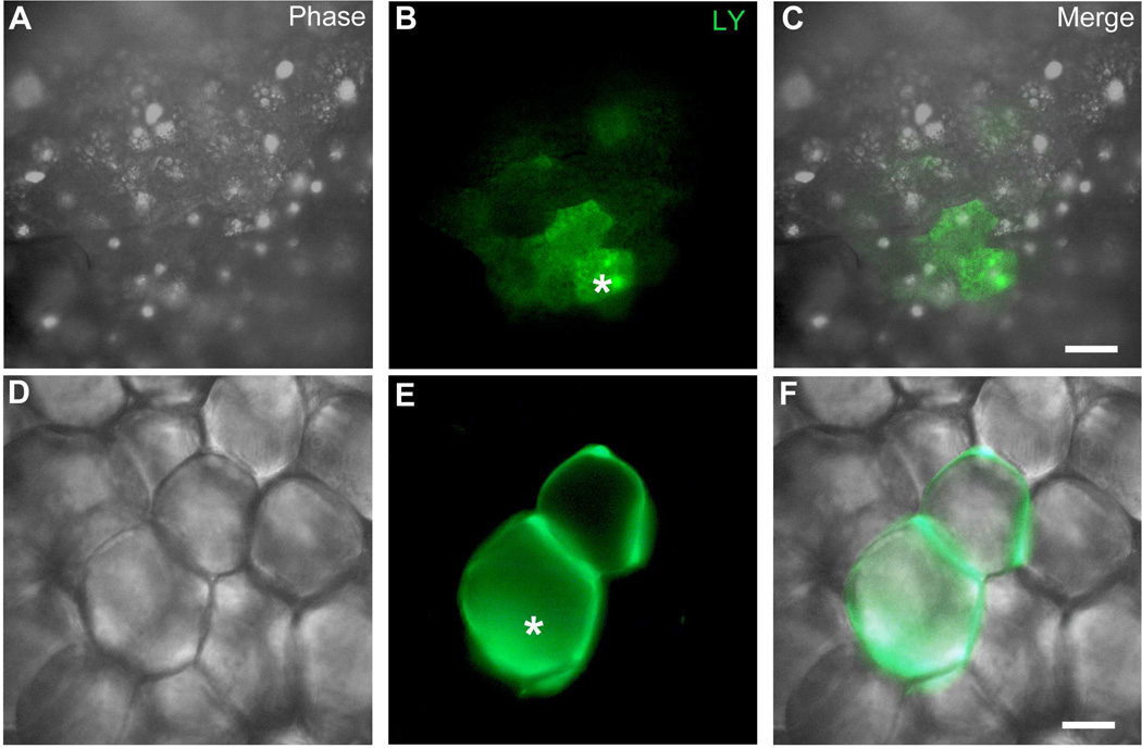
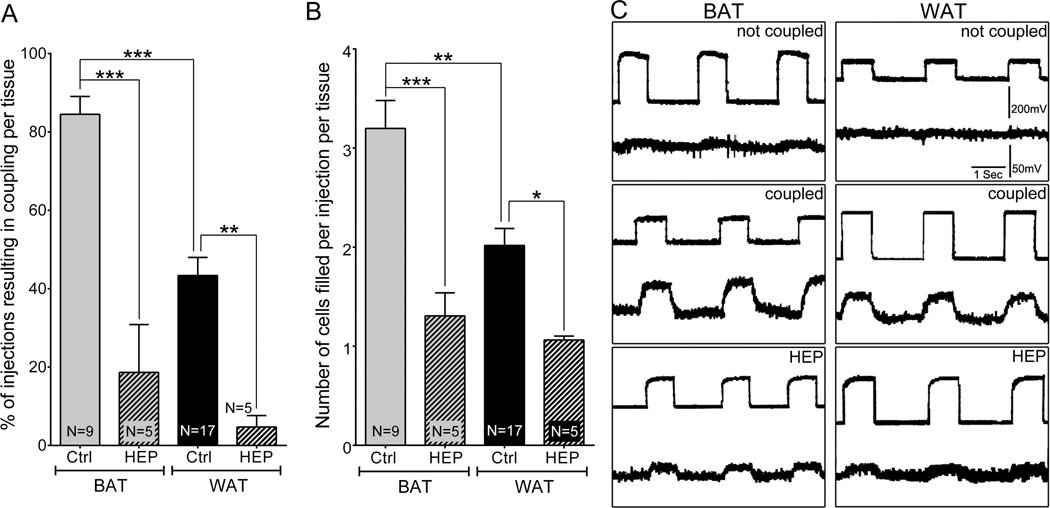
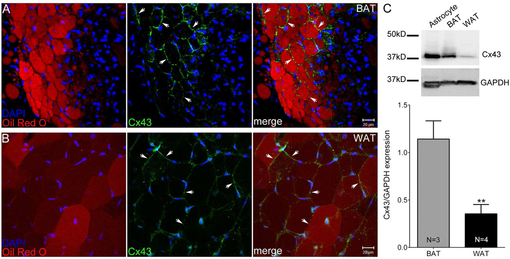
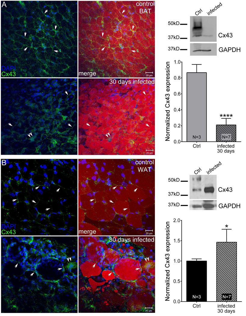
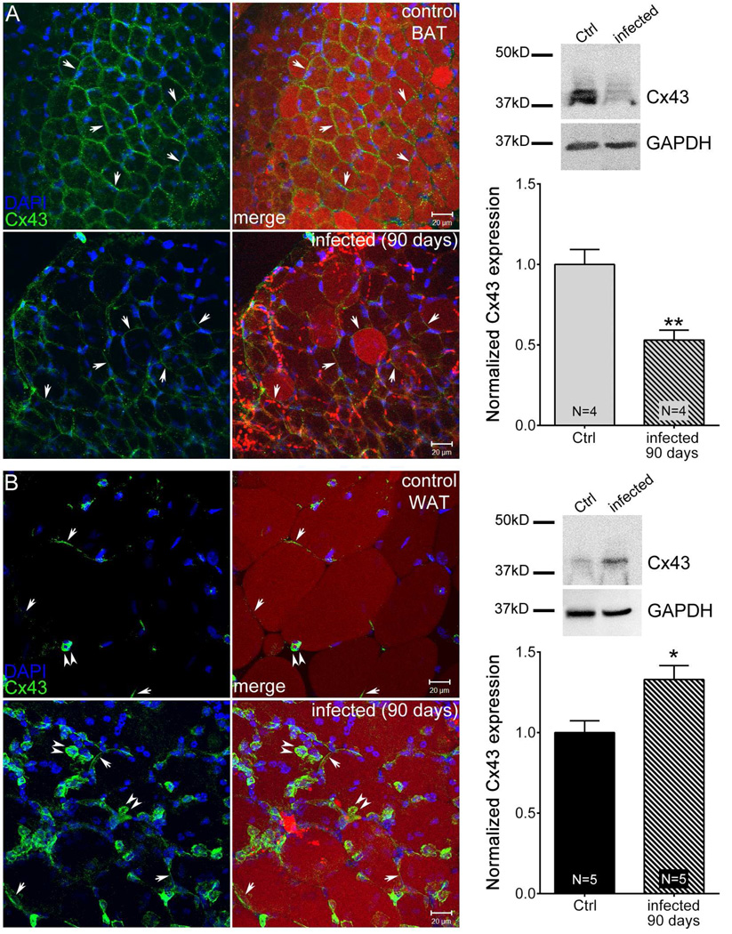
Adipose tissue serves as a host reservoir for the protozoan Trypanosoma cruzi, the causative organism in Chagas disease. Gap junctions interconnect cells of most tissues, serving to synchronize cell activities including secretion in glandular tissue, and we have previously demonstrated that gap junctions are altered in various tissues and cells infected with T. cruzi. Herein, we examined the gap junction protein connexin 43 (Cx43) expression in infected adipose tissues. Adipose tissue is the largest endocrine organ of the body and is also involved in other physiological functions. In mammals, it is primarily composed of white adipocytes. Although gap junctions are a prominent feature of brown adipocytes, they have not been explored extensively in white adipocytes, especially in the setting of infection. Thus, we examined functional coupling in both white and brown adipocytes in mice. Injection of electrical current or the dye Lucifer Yellow into adipocytes within fat tissue spread to adjacent cells, which was reduced by treatment with agents known to block gap junctions. Moreover, Cx43 was detected in both brown and white fat tissue. At thirty and ninety days post-infection, Cx43 was downregulated in brown adipocytes and upregulated in white adipocytes. Gap junction-mediated intercellular communication likely contributes to hormone secretion and other functions in white adipose tissue and to nonshivering thermogenesis in brown fat, and modulation of the coupling by T. cruzi infection is expected to impact these functions.

Keywords: Adipose tissue; Chagas disease; Connexin43; Gap junctions; Trypanosoma cruzi.


Figures

Figure 1
Figure 1
Functional coupling in adipose tissue. A–F. Dye injection. Individual adipocytes were intracellularly injected with the fluorescent dye Lucifer yellow (LY). Asterisk indicates the injected cell. A–C: Brown adipose tissue (BAT) with dye coupling to neighboring cells; D–F: White adipose tissue (WAT) with dye spread to one neighboring cell. Scale bar = 30 µm.
Figure 2
Figure 2
Quantification of dye coupling and its blockade by the gap junction inhibitor heptanol. A: Graph showing the mean percentage of injections per tissue that resulted in coupling. B: Graph showing the mean number of cells seen in each injection per tissue sample. All data are presented as mean ± SEM, N ≥ 5. P values were obtained using one way ANOVA analysis followed by Tukey’s multiple comparison test. (*P < 0.05, **P < 0.005, ***P < 0.0005, control vs. treated). C: Electrical coupling in brown and white adipose tissue. Current pulses were delivered into one cell and voltage deflections recorded in that cell (top) and a neighbor (bottom trace) shown by LY to be coupled to the first, injected cell. Lack of measureable current spread from electrode within the cell (upper trace) and an extracellular electrode before impalement of a second cell (not coupled), measureable spread of current when the second microelectrode was placed into a coupled cell (coupled), and significant reduction in current when tissue with the second electrode still in the same coupled cell, after the sample was incubated for 1 h with 3mM heptanol (HEP).
Figure 3
Figure 3
Biochemical evidence for Cx43 in adipose tissue. Confocal image stacks showing Cx43 distribution (green, arrows) together with Oil-Red-O (red) and DAPI (blue) staining in (A) Brown adipose tissue (BAT) and (B) white adipose tissue (WAT). Scale bar = 20 µm. C. Representative Western blots of Cx43 and GAPDH in BAT and WAT and quantification of Cx43 levels normalized with respect to internal loading control GAPDH. All data are presented as mean ± SEM, (N = 3 [BAT], N = 4 [WAT]). P value was obtained using unpaired t-test. (**P < 0.005, BAT vs. WAT)
Figure 4
Figure 4
Acute T.cruzi infection alters Cx43 distribution and expression in adipose tissue. Animals were infected with 5×10⁴ Brazilian Strain T. cruzi for 30 days. Confocal image stacks from control brown adipose tissue (BAT) (A, upper panel), infected BAT (A, lower panel), control white adipose tissue (WAT) (B, upper panel) and infected WAT (B, lower panel) showing Cx43 distribution (green, arrows), Oil-Red-O (red) and DAPI (blue) staining. Scale bar = 20 µm. Representative Western blots and normalized expression of Cx43 from 30 day infected mice (BAT [A] and WAT [B]) compared to that of respective control. The blots were analyzed using image J and results were normalized first to GAPDH followed by control. All data are presented as mean ± SEM. P values were obtained using unpaired t-test. (**P < 0.005, ****P < 0.00005, Control vs. infected adipose tissue)
Figure 5
Figure 5
Chronic T. cruzi infection alters Cx43 distribution and expression in adipose tissue. Animals were infected with 5×10⁴ Brazilian Strain T. cruzi for 90 days. Stacked confocal images from control BAT (A, upper panel), infected BAT (A, lower panel), control white adipose tissue (WAT) (B, upper panel) and infected WAT (B, lower panel) showing Cx43 distribution (green, arrows), Oil-Red-O (red) and DAPI (blue) staining. Scale bar = 20 µm. Representative Western blots and normalized expression of Cx43 from 30 day infected mice (BAT [A] and WAT [B]) compared to that of respective control. The blots were analyzed using image J and results were normalized first to GAPDH followed by control. All data are presented as mean ± SEM. P values were obtained using unpaired t-test. (*P < 0.05, **P < 0.005, Control vs. infected adipose tissue)

References

    1. Spray DC, Hanstein R, Lopez-Quintero SV, Stout RF, Jr, Suadicani SO, Thi MM. Gap junctions and Bystander Effects: Good Samaritans and executioners. Wiley Interdiscip Rev Membr Transp Signal. 2013;2:1–15. - PMC - PubMed
    1. Michon L, Nlend Nlend R, Bavamian S, Bischoff L, Boucard N, Caille D, Cancela J, Charollais A, Charpantier E, Klee P, Peyrou M, Populaire C, Zulianello L, Meda P. Involvement of gap junctional communication in secretion. Biochim Biophys Acta. 2005;1719:82–101. - PubMed
    1. Halberg N, Wernstedt-Asterholm I, Scherer PE. The adipocyte as an endocrine cell. Endocrinol Metab Clin North Am. 2008;37:753–768. x–xi. - PMC - PubMed
    1. Scherer PE. Adipose tissue: from lipid storage compartment to endocrine organ. Diabetes. 2006;55:1537–1545. - PubMed
    1. Nedergaard J, Bengtsson T, Cannon B. Unexpected evidence for active brown adipose tissue in adult humans. Am J Physiol Endocrinol Metab. 2007;293:E444–E452. - PubMed

Publication types