. 2014 Oct 10;173(3-4):177-88.
doi: 10.1016/j.vetmic.2014.07.020. Epub 2014 Aug 4.

Mutations of 3c and spike protein genes correlate with the occurrence of feline infectious peritonitis

Affiliations

Mutations of 3c and spike protein genes correlate with the occurrence of feline infectious peritonitis

Barbara Regina Bank-Wolf et al. Vet Microbiol. .

Abstract

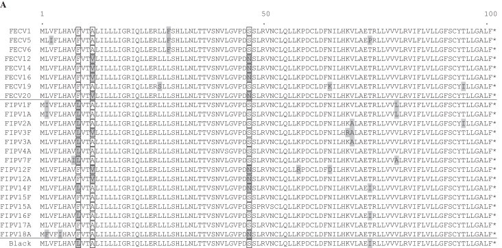
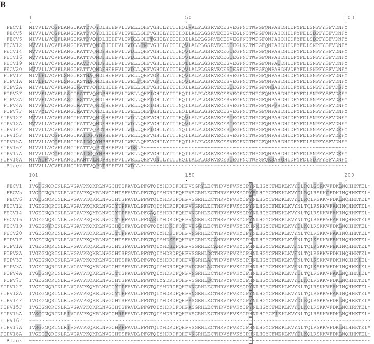
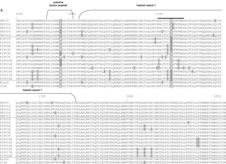
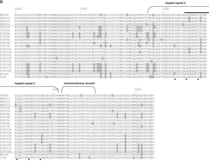
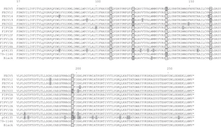
The genes encoding accessory proteins 3a, 3b, 3c, 7a and 7b, the S2 domain of the spike (S) protein gene and the membrane (M) protein gene of feline infectious peritonitis virus (FIPV) and feline enteric coronavirus (FECV) samples were amplified, cloned and sequenced. For this faeces and/or ascites samples from 19 cats suffering from feline infectious peritonitis (FIP) as well as from 20 FECV-infected healthy cats were used. Sequence comparisons revealed that 3c genes of animals with FIP were heavily affected by nucleotide deletions and point mutations compared to animals infected with FECV; these alterations resulted either in early termination or destruction of the translation initiation codon. Two ascites-derived samples of cats with FIP which displayed no alterations of ORF3c harboured mutations in the S2 domain of the S protein gene which resulted in amino acid exchanges or deletions. Moreover, changes in 3c were often accompanied by mutations in S2. In contrast, in samples obtained from faeces of healthy cats, the ORF3c was never affected by such mutations. Similarly ORF3c from faecal samples of the cats with FIP was mostly intact and showed only in a few cases the same mutations found in the respective ascites samples. The genes encoding 3a, 3b, 7a and 7b displayed no mutations linked to the feline coronavirus (FCoV) biotype. The M protein gene was found to be conserved between FECV and FIPV samples. Our findings suggest that mutations of 3c and spike protein genes correlate with the occurrence of FIP.

Keywords: Accessory genes; FECV; FIPV; Feline coronavirus; Spike protein gene.


Figures

Fig. 1
Fig. 1
Sequence alignments of accessory proteins 7a (A) and 7b (B). Complete 7a comprises 101 aa, complete 7b comprises 206 aa residues. Residues diverging from the consensus are highlighted by grey boxes. For 7a, aa at positions 9, 12 and 47 and for 7b at position 170 are highlighted by translucent boxes (see text). 7b of FIPV-Black and FIPV-16F is truncated to 35 residues. Accession number of reference strain Black: EU186072.
Fig. 1
Fig. 1
Sequence alignments of accessory proteins 7a (A) and 7b (B). Complete 7a comprises 101 aa, complete 7b comprises 206 aa residues. Residues diverging from the consensus are highlighted by grey boxes. For 7a, aa at positions 9, 12 and 47 and for 7b at position 170 are highlighted by translucent boxes (see text). 7b of FIPV-Black and FIPV-16F is truncated to 35 residues. Accession number of reference strain Black: EU186072.
Fig. 2
Fig. 2
Sequence alignment of the putative fusion peptide and adjacent heptad repeat 1 (HR1) domain (A) and a part of the linker region and adjacent heptad repeat 2 (HR2) (B) of the spike protein. Residues diverging from the consensus are highlighted by grey boxes; aa at positions 1045 and 1108 are highlighted by translucent boxes. The putative fusion peptide and HR 1 and HR 2 domains are indicated by brackets. The leucine zipper domain in HR 2 is highlighted by black dots. A stretch of 14 aa comprising exactly two heptad repeats which is only found in alphacoronaviruses is indicated by a black bar. Numbers refer to amino acid position in spike protein of strain Black. Accession numbers of reference strains: Black: EU186072, C1Je: dq848678. Amino acids EY at position 1217/1218 of FECV were replaced with DH or QY in most FIPV spike proteins. Further noticeable changes were D1210N, E1267D/Q, Y1268H, N1275G, F1295L, T1299A/S, T1308A, YTT/I to FV/SN/R/M (1310–1312), E1315D/N, Y1334H, T/AP to I/TS/T (1341/1342), N/H/DL to HF/L (1345/1346), H1444N and S1454R/N (B).
Fig. 2
Fig. 2
Sequence alignment of the putative fusion peptide and adjacent heptad repeat 1 (HR1) domain (A) and a part of the linker region and adjacent heptad repeat 2 (HR2) (B) of the spike protein. Residues diverging from the consensus are highlighted by grey boxes; aa at positions 1045 and 1108 are highlighted by translucent boxes. The putative fusion peptide and HR 1 and HR 2 domains are indicated by brackets. The leucine zipper domain in HR 2 is highlighted by black dots. A stretch of 14 aa comprising exactly two heptad repeats which is only found in alphacoronaviruses is indicated by a black bar. Numbers refer to amino acid position in spike protein of strain Black. Accession numbers of reference strains: Black: EU186072, C1Je: dq848678. Amino acids EY at position 1217/1218 of FECV were replaced with DH or QY in most FIPV spike proteins. Further noticeable changes were D1210N, E1267D/Q, Y1268H, N1275G, F1295L, T1299A/S, T1308A, YTT/I to FV/SN/R/M (1310–1312), E1315D/N, Y1334H, T/AP to I/TS/T (1341/1342), N/H/DL to HF/L (1345/1346), H1444N and S1454R/N (B).
Fig. 3
Fig. 3
Sequence alignment of aa 57–263 of the membrane protein. Residues diverging from the consensus are highlighted by grey boxes. Numbers refer to amino acid position in spike protein of strain Black. Clear boxes indicate five aa positions formerly described as being marker mutations. Accession numbers of reference strains: p04135: TGEV, 79–1146 (serotype II): AY994055, Black (serotype I): EU186072.

References

    1. Becher P., Orlich M., Shannon A.D., Horner G., Konig M., Thiel H.J. Phylogenetic analysis of pestiviruses from domestic and wild ruminants. J. Gen. Virol. 1997;78(Pt 6):1357–1366. - PubMed
    1. Brown M.A. Genetic determinants of pathogenesis by feline infectious peritonitis virus. Vet. Immunol. Immunopathol. 2011;143(3/4):265–268. - PMC - PubMed
    1. Brown M.A., Troyer J.L., Pecon-Slattery J., Roelke M.E., O’Brien S.J. Genetics and pathogenesis of feline infectious peritonitis virus. Emerg. Infect. Dis. 2009;15(9):1445–1452. - PMC - PubMed
    1. Chang H.W., de Groot R.J., Egberink H.F., Rottier P.J. Feline infectious peritonitis: insights into feline coronavirus pathobiogenesis and epidemiology based on genetic analysis of the viral 3c gene. J. Gen. Virol. 2010;91(Pt 2):415–420. - PubMed
    1. Chang H.W., Egberink H.F., Halpin R., Spiro D.J., Rottier P.J. Spike protein fusion peptide and feline coronavirus virulence. Emerg. Infect. Dis. 2012;18(7):1089–1095. - PMC - PubMed

Publication types

MeSH terms

LinkOut - more resources