

Non-uniform displacements within the Achilles tendon observed during passive and eccentric loading
- PMID: 25150898
- PMCID: PMC4163107
- DOI: 10.1016/j.jbiomech.2014.07.032
Non-uniform displacements within the Achilles tendon observed during passive and eccentric loading
Abstract
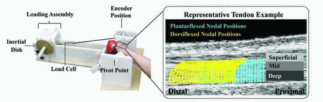
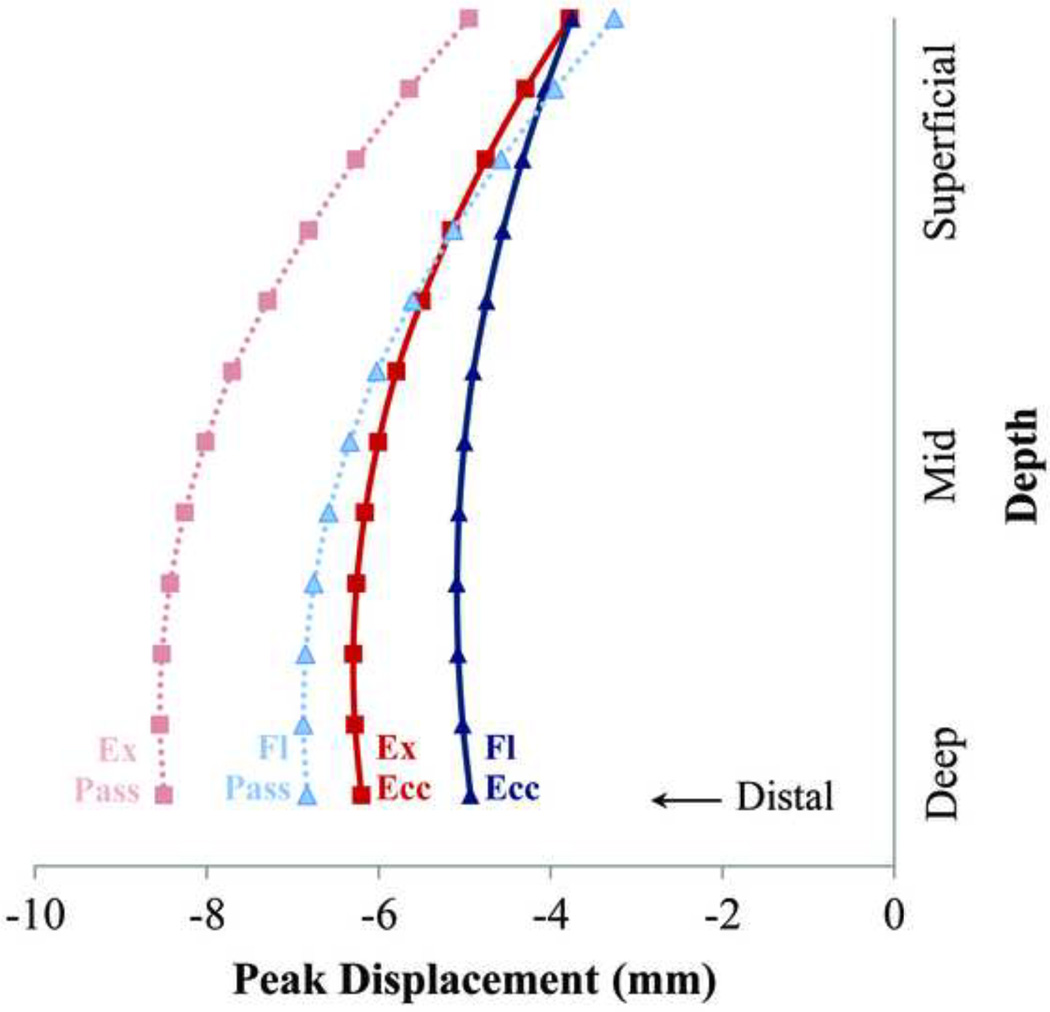
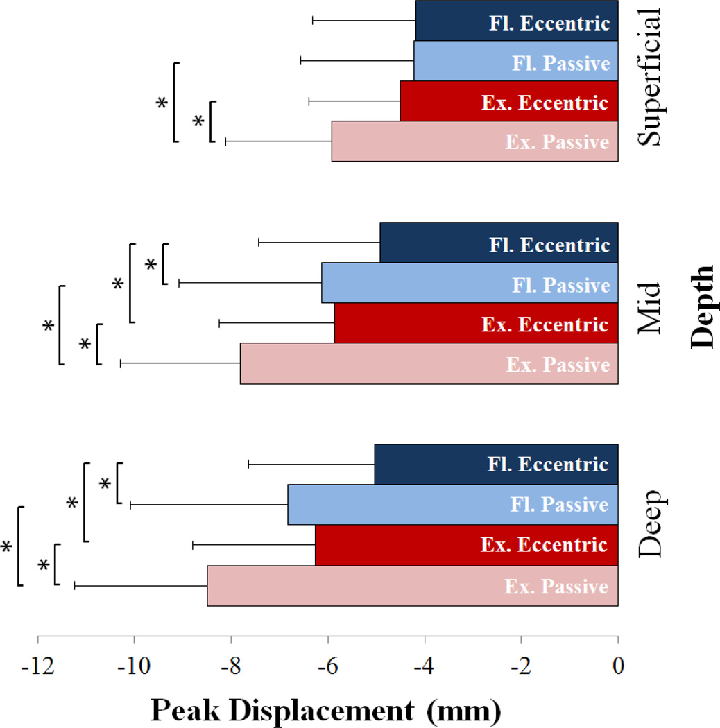
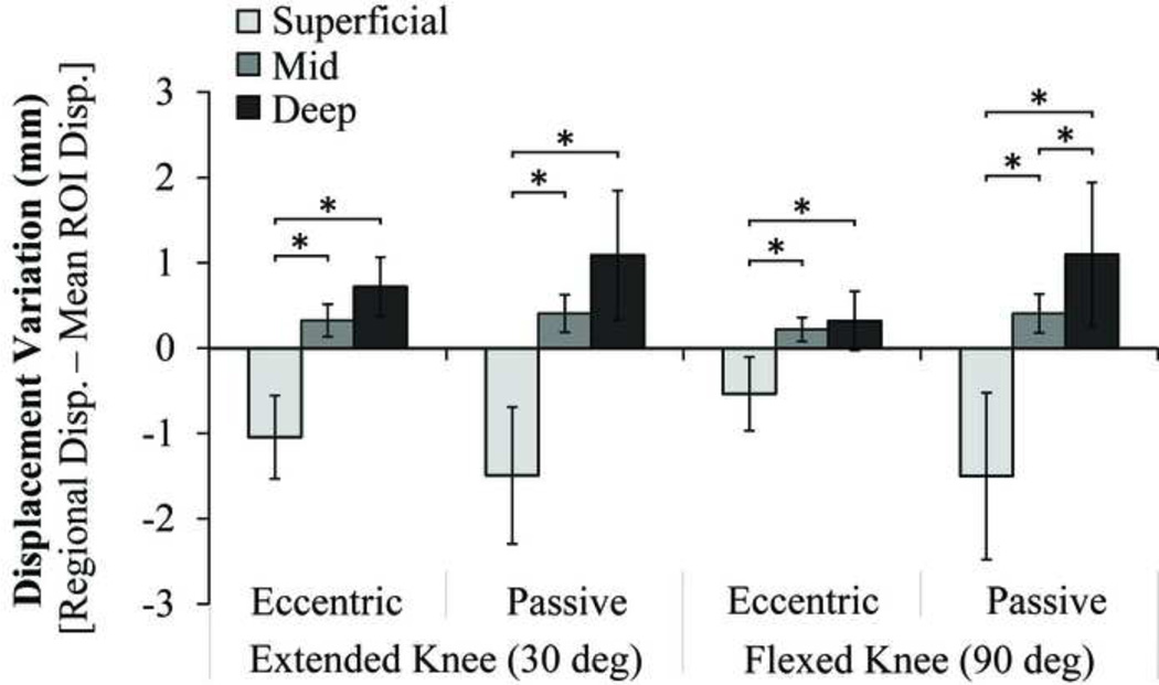
The goal of this study was to investigate Achilles tendon tissue displacement patterns under passive and eccentric loading conditions. Nine healthy young adults were positioned prone on an examination table with their foot secured to a rotating footplate aligned with the ankle. Subjects cyclically rotated their ankle over a 25° range of motion at 0.5 Hz. An inertial load geared to the footplate induced eccentric plantarflexor contractions with dorsiflexion. Passive cyclic ankle motion was also performed over the same angular range of motion. An ultrasound transducer positioned over the distal Achilles tendon was used to collect radiofrequency (RF) images at 70 frames/s. Two-dimensional ultrasound elastographic analysis of the RF data was used to track tendon tissue displacements throughout the cyclic motion. Non-uniform tissue displacement patterns were observed in all trials, with the deeper portions of the Achilles tendon consistently exhibiting larger displacements than the superficial tendon (average of 0.9-2.6mm larger). Relative to the passive condition, eccentric loading consistently induced smaller tissue displacements in all tendon regions, except for the superficial tendon in a flexed knee posture. Significantly greater overall tissue displacement was observed in a more extended knee posture (30°) relative to a flexed knee posture (90°). These spatial- and posture-dependent displacement patterns suggest that the tendon undergoes non-uniform deformation under in vivo loading conditions. Such behavior could reflect relative sliding between the distinct tendon fascicles that arise from the gastrocnemius and soleus muscles.
Keywords: Achilles tendon; Non-uniform motion; Ultrasound elastography.
Copyright © 2014 Elsevier Ltd. All rights reserved.
Conflict of interest statement
Conflict of interest statement
The authors have no conflict of interest.
Figures
References
-
- Alfredson H. Chronic midportion Achilles tendinopathy: an update on research and treatment. Clin. Sports Med. 2003;22:727–741. - PubMed
-
- Arampatzis A, Karamanidis K, Stafilidis S, Morey-Klapsing G, DeMonte G, Bruggemann GP. Effect of different ankle- and knee-joint positions on gastrocnemius medialis fascicle length and EMG activity during isometric plantar flexion. J. Biomech. 2006;39:1891–1902. - PubMed
-
- Arampatzis A, Stafilidis S, DeMonte G, Karamanidis K, Morey-Klapsing G, Bruggenmann GP. Strain and elongation of the human gastrocnemius tendon and aponeurosis during maximal plantarflexion effort. J. Biomech. 2005;38:833–841. - PubMed
-
- Arndt A, Bengtsson AS, Peolsson M, Thorstensson A, Movin T. Non-uniform displacement within the Achilles tendon during passive ankle joint motion. Knee surgery, sports traumatology, arthroscopy: official journal of the ESSKA. 2012;20:1868–1874. - PubMed
-
- Arndt A, Bruggemann GP, Koebke J, Segesser B. Asymmetrical loading of the human triceps surae: I. Mediolateral force differences in the Achilles tendon. Foot Ankle Int. 1999;20:444–449. - PubMed
Publication types
MeSH terms
Grants and funding
LinkOut - more resources
Full Text Sources
Other Literature Sources
