Review
. 2014 Nov;17(11):1500-9.
doi: 10.1038/nn.3776. Epub 2014 Aug 24.

Dimensionality reduction for large-scale neural recordings

Affiliations
Review

Dimensionality reduction for large-scale neural recordings

John P Cunningham et al. Nat Neurosci. 2014 Nov.

Abstract

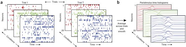
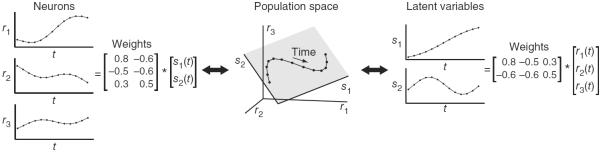
Most sensory, cognitive and motor functions depend on the interactions of many neurons. In recent years, there has been rapid development and increasing use of technologies for recording from large numbers of neurons, either sequentially or simultaneously. A key question is what scientific insight can be gained by studying a population of recorded neurons beyond studying each neuron individually. Here, we examine three important motivations for population studies: single-trial hypotheses requiring statistical power, hypotheses of population response structure and exploratory analyses of large data sets. Many recent studies have adopted dimensionality reduction to analyze these populations and to find features that are not apparent at the level of individual neurons. We describe the dimensionality reduction methods commonly applied to population activity and offer practical advice about selecting methods and interpreting their outputs. This review is intended for experimental and computational researchers who seek to understand the role dimensionality reduction has had and can have in systems neuroscience, and who seek to apply these methods to their own data.


Figures

Figure 1
Figure 1
Motivation for population analyses and dimensionality reduction. (a) Enabled by the growth in scale and resolution of neural recording technologies, a typical experiment yields a collection of many trials (sets of panels from left to right), many experimental conditions (different colored panels shown in depth) and many neurons (rows of each panel, shown as spike rasters). Scrutinizing these data qualitatively and quantitatively presents many challenges, both for basic understanding and for testing hypotheses. (b) Neural responses are often averaged across trials (within a given condition) and smoothed into a peristimulus time histogram. Even these trial-averaged views can be difficult to interpret, as the number of conditions and recorded neurons grows. Notably, this challenge can be present even in data with simple structure: each of these simulated neurons have Poisson spiking with an underlying firing rate that is a windowed linear mixture of three Gaussian pulses. Each neuron has different mixture coefficients, baseline and amplitude. Dimensionality reduction is one class of statistical methods that can extract simple structure from these seemingly complex data.
Figure 2
Figure 2
Conceptual illustration of linear dimensionality reduction for three neurons (D = 3) and two latent variables (K = 2). Center, the population activity (black points) lies in a plane (shaded gray). Each point represents the population activity at a particular time and can be equivalently referred to using its high-dimensional coordinates [r1, r2, r3] or low-dimensional coordinates [s1, s2]. The points trace out a trajectory over time (black curve). Left, the population activity r1, r2 and r3 can be reconstructed by taking a weighted combination of the latent variables, where the weights are specified by the matrix shown. Right, the latent variables s1 and s2 can be obtained by taking a weighted combination of the population activity, where the weights are specified by the matrix shown.
Figure 3
Figure 3
Examples of scientific studies using dimensionality reduction. (a) Single-trial statistical power, visual attention. Left, a didactic example of projecting the responses of two neurons onto the attention axis (green; units, spike counts). For a V4 population (right), the normalized position (that is, projection) along this attention axis was predictive of behavior on single trials: the farther the projection was to the upper right of the attention axis, the more likely the animal was of correctly detecting right changes and the less likely the animal was of correctly detecting left changes. Adapted with permission from ref. 17. (b) Population response structure, decision-making. The population activity recorded in prefrontal cortex was projected onto three axes (units, spikes per s): the axis of evidence integration (choice axis), the relevant stimulus axis (motion axis) and the irrelevant stimulus axis (color axis). Each trace corresponds to responses averaged across trials of the same dot motion (gray traces) or dot color (blue traces). Despite the apparent complexity of single-neuron responses, the population activity shows orderly structure across different conditions of dot motion and color and suggests a network mechanism for gating and integration of information in prefrontal cortex. Adapted with permission from ref. 4. (c) Population response structure, motor system. The population activity recorded in motor cortex was projected onto a plane (units, spikes per s) where simple (rotational) dynamics are best captured; different traces are different experimental conditions (arm reaches shown with the same color in the inset). Dots denote the preparatory (pre-movement) neural activity, suggesting a mechanistic explanation for single neuron complexity: preparatory responses set the initial state of a population-level dynamical system that runs through movement. Adapted with permission from ref. 11. (d) Exploratory data analysis, brain-wide. The population activity recorded throughout the brain of larval zebrafish was projected onto its principal components for visualization purposes (the same data is shown at left plotted in three and two dimensions, units of ΔF/F). Four phases of response were identified (labeled α, β, γ and δ), which were then connected back to distinct neural structures. Right, axial views during each phase, where green dots indicate active neurons with magenta confidence intervals (caudal-rostral is left-right). Adapted with permission from ref. 50.
Figure 4
Figure 4
Conceptual illustration of PCA, LDA and demixed dimensionality reduction for two neurons (D = 2). (a) PCA finds the direction (s1 axis) that captures the greatest variance in the data (black dots, top), shown by the projection onto the s1 axis (bottom). (b) LDA finds the direction (s1 axis) that best separates the two groups of points (black and while dots, top). The separation can be seen in the projection onto the s1 axis (bottom). (c) Demixed dimensionality reduction (using the method described in ref. 16 finds the direction that explains the variance in dot color (s1 axis, top) and an orthogonal direction (s2 axis, not shown) that explains the variance in dot size. The organization in dot color can be seen in the projection onto the s1 axis (bottom). Note that these illustrations were created using the same data points (dots), and it is the use of different methods (which exploit different data features, such as group membership in (b) or color and size in (c)) that produce different directions s1 across the top panels and different projections across the bottom panels.

References

    1. Kipke DR, et al. Advanced neurotechnologies for chronic neural interfaces: new horizons and clinical opportunities. J. Neurosci. 2008;28:11830–11838. - PMC - PubMed
    1. Kerr JN, Denk W. Imaging in vivo: watching the brain in action. Nat. Rev. Neurosci. 2008;9:195–205. - PubMed
    1. Ahrens MB, et al. Whole-brain functional imaging at cellular resolution using light-sheet microscopy. Nat. Methods. 2013;10:413–420. - PubMed
    1. Mante V, et al. Context-dependent computation by recurrent dynamics in prefrontal cortex. Nature. 2013;503:78–84. - PMC - PubMed
    1. Kaufman MT, et al. Cortical activity in the null space: permitting preparation without movement. Nat. Neurosci. 2014;17:440–448. - PMC - PubMed

Publication types

LinkOut - more resources