. 2014 Oct;15(10):965-72.
doi: 10.1038/ni.2981. Epub 2014 Aug 24.

The kinase p38 activated by the metabolic regulator AMPK and scaffold TAB1 drives the senescence of human T cells

Affiliations

The kinase p38 activated by the metabolic regulator AMPK and scaffold TAB1 drives the senescence of human T cells

Alessio Lanna et al. Nat Immunol. 2014 Oct.

Abstract

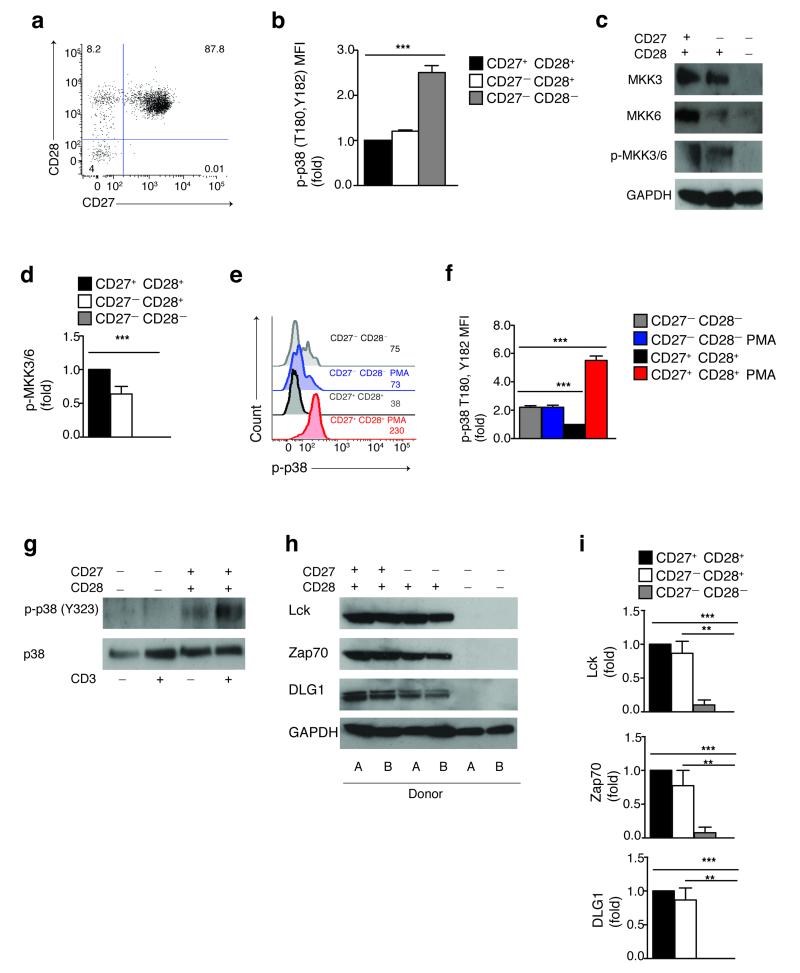
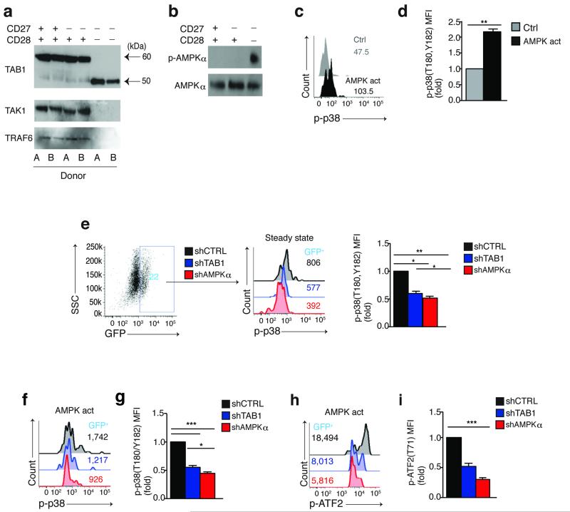
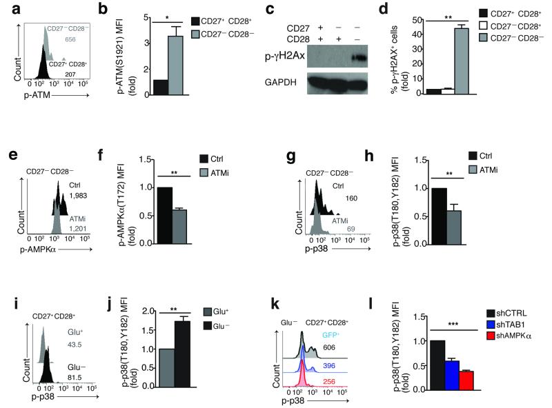
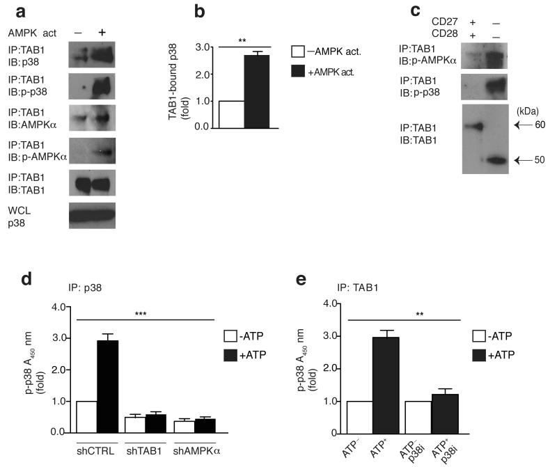
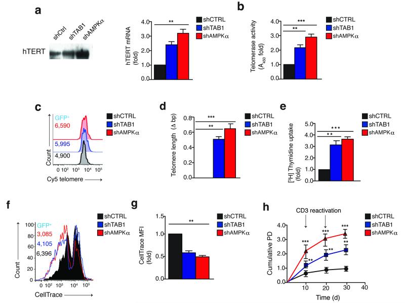
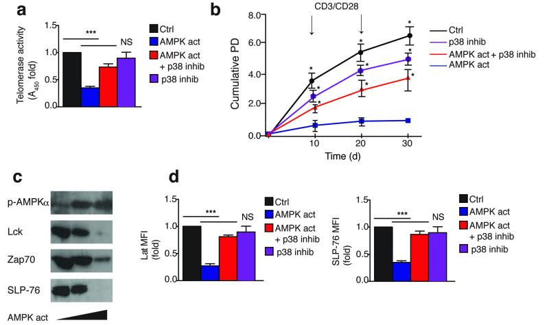
In T lymphocytes, the mitogen-activated protein kinase (MAPK) p38 regulates pleiotropic functions and is activated by canonical MAPK signaling or the alternative activation pathway downstream of the T cell antigen receptor (TCR). Here we found that senescent human T cells lacked the canonical and alternative pathways for the activation of p38 but spontaneously engaged the metabolic master regulator AMPK to trigger recruitment of p38 to the scaffold protein TAB1, which caused autophosphorylation of p38. Signaling via this pathway inhibited telomerase activity, T cell proliferation and the expression of key components of the TCR signalosome. Our findings identify a previously unrecognized mode for the activation of p38 in T cells driven by intracellular changes such as low-nutrient and DNA-damage signaling (an 'intrasensory' pathway). The proliferative defect of senescent T cells was reversed by blockade of AMPK-TAB1-dependent activation of p38.


Figures

Figure 1
Figure 1. Spontaneous p38 activation in the absence of both upstream canonical and alternative pathways in highly differentiated T cells
(a) The co-expression of CD27 and CD28 receptors by human CD4+ T cells, representative staining (n=9). (b) Phospho-flow data of spontaneous p38 (Thr180,Tyr182) phosphorylation in CD4+ CD27/CD28 defined subsets from 9 different donors (MFI, mean fluorescence intensity). (c) Immunoblots of total MKK3, MKK6 and phospho-MKK3/MKK6 (Ser189,Ser206) in CD4+ CD27/CD28 defined subsets. GAPDH was used as a loading control. Data are representative of 4 independent experiments. (d) The relative level of endogenous MKK3/6 activation vs. total GAPDH in CD4+ CD27/CD28 defined subsets of 4 separate individuals. (e) Representative overlay and (f) pooled phospho-flow data from 3 independent experiments showing p38 (Thr180,Tyr182) phosphorylation in CD27 CD28 and CD27+ CD28+ CD4+ T cells either before or after treatment with PMA (20 ng/mL, 60′). (g) Immunoblots of total p38 and phospho-p38 (Tyr323) in isolated CD27 CD28 and CD27+ CD28+ CD4+ T cells either before or after αCD3 activation (10 μg/mL, 30′). Data are representative of 3 independent experiments. (h) Representative blots of total Lck, Zap70 and DLG1 expression in CD4+ CD27/CD28 defined subsets; GAPDH was used as a loading control. Two different donors from the same gel are shown. (i) The relative level of Lck, Zap70 and DLG1 protein expression vs. total GAPDH in CD4+ CD27/CD28 defined subsets of 4 different subjects. All *p< 0,05, **p<0.01, and ***p< 0.001 values were calculated using one-way analysis of variance (ANOVA) for repeated-measures with a Bonferroni post-test correction. Error bars depict s.e.m.
Figure 2
Figure 2. Spontaneous p38 activation triggered by AMPK and mediated by TAB1 in highly differentiated T cells
(a) Immunoblots of TAB1 (alternative isoforms depicted by black arrows), TAK1 and TRAF6 expression from 2 different subjects within the same gel and (b) total AMPKα and phospho-AMPKα (Thr172) expression in freshly isolated CD4+ CD27/CD28 defined subsets. Data are representative of (a) 4 or (b) 3 different donors. (c) Representative overlay and (d) pooled data from 3 separate experiments showing the effect of the AMPK activator A-769662 (150 μM, 60′) on p38 (Thr180,Tyr182) phosphorylation in CD27+ CD28+ CD4+ T cells as determined by phospho-flow analysis. (e) Representative FACS plot (n=9) showing expression of the reporter GFP gene in transduced purified CD27CD28 CD4+ T cells, 96 hours post transduction (left); overlay from the same experiment (middle) and pooled phospho-flow data from 3 separate experiments (right) showing steady-state p38 (Thr180,Tyr182) phosphorylation within the reporter GFP+ populations of CD27CD28 CD4+ T cells transduced with lentiviral vectors encoding either shAMPKα, shTAB1 or shCtrl (knock-down validation in Supplementary Fig. 2). (f) Representative overlay and (g) pooled data from 3 separate experiments showing the effect of the AMPK activator A-769662 (150 μM, 60′) on p38 (Thr180,Tyr182) phosphorylation in CD27 CD28 CD4+ T cells transduced as indicated and analyzed within the GFP+ subsets by phospho-flow. (h) Representative overlay and (i) pooled phospho-flow data of ATF2 (Thr71) phosphorylation from 3 separate experiments performed as described in (f,g). In (d) a paired Student’s t test was used. For all other statistics a one-way analysis of variance (ANOVA) for repeated-measures with a Bonferroni post-test correction. p< 0,05, **p<0.01, and ***p< 0.001. Error bars depict s.e.m.
Figure 3
Figure 3. DNA damage and low-nutrient signals converge at AMPK which drives TAB1 dependent p38 activation in T cells
(a) Representative overlay and (b) pooled phospho-flow data from 4 different subjects of spontaneous ATM (Ser1921) phosphorylation in CD27CD28 and CD27+ CD28+ CD4+ T cells, directly ex vivo. (c) Immunoblot and (d) pooled phospho-flow data from 4 different subjects of γH2AX (Ser139) phosphorylation in freshly isolated CD4+ CD27/CD28 defined subsets; GAPDH served as loading control in (c). Immunoblot is representative of 2 independent experiments. (e) Representative overlay and (f) pooled phospho-flow data showing the effect of the selective ATM inhibitor KU-55933 (60′, 10 μM) on constitutive AMPKα (Thr172) phosphorylation in CD27 CD28 CD4+ T cells. (g) Representative overlay and (h) pooled phospho-flow data of p38 (Thr180,Tyr182) phosphorylation from 3 separate experiments performed as described in (e,f). (i) Representative overlay and (j) pooled results from 3 separate experiments showing the effect of 18 hours of glucose starvation on p38 (Thr180,Tyr182) phosphorylation in CD27+ CD28+ CD4+ T cells. (k) Representative overlay and (l) pooled data showing the effect of 18 hours glucose starvation on p38 (Thr180,Tyr182) phosphorylation in CD27+ CD28+ CD4+ T cells transduced as indicated and analyzed within the GFP+ subsets by phospho-flow.
Figure 4
Figure 4. AMPK triggered p38 recruitment to TAB1 causes p38 auto-phosphorylation
(a) Immunoblots of total p38, phospho-p38 (Thr180,Tyr182), total AMPKα phospho-AMPKα (Thr172) and TAB1 in CD27+ CD28+ CD4+ T cells activated with DMSO control or the AMPK agonist A-769662 (150 μM) for 2 hours, followed by immunoprecipitation with anti-TAB1. Data are representative of 4 separate donors. Immunoblot of total p38 from whole cell lysate (WCL) served as input control. (b) The relative binding of p38 to TAB1 upon AMPK activation as determined by 4 independent experiments performed as described in (a). (c) Freshly-isolated CD27+ CD28+ or CD27 CD28 CD4+ T cells were immunoprecipitated with anti-TAB1 and analyzed by immunoblot, as indicated (alternative TAB1 isoforms depicted by black arrows). Data are representative of 2 separate experiments (for the minor CD4+ CD27 CD28 T cell fraction, cells were pooled together from 2 different donors to achieve sufficient cell number to perform the assay). (d) Measurement of p38 auto-phosphorylation by ELISA-based in vitro kinase assay of p38 immunoprecipitates from transduced purified CD27+ CD28+ CD4+ T cells reactivated with the AMPK agonist A-769662 (150 μM) for 2 hours. Immunoprecipitates were left untreated or incubated for 30 min with ATP (200 μM). (e) Measurement of p38 auto-phosphorylation of TAB1 immunoprecipitates from CD4+ CD27+ CD28+ T cells activated with the AMPK agonist A-769662 (150 μM) for 2 hours. The assay was performed as described in (d) in the presence or absence of the p38 inhibitor SB-203580 (10 μM). Experiments in (d,e) were perfomed from 3 different donors. In (b) a paired Student’s t test was used; for (d) and (e) a one-way analysis of variance (ANOVA) for repeated-measures with a Bonferroni post-test correction. *p< 0,05, **p<0.01, and ***p< 0.001. Error bars depict s.e.m.
Figure 5
Figure 5. Silencing of AMPK or TAB1 restores telomerase and proliferation in highly differentiated T cells
(a) Measurement of hTERT expression by both immunoblot (left) and quantitative PCR (right) in CD27 CD28 CD4+ T cells, transduced as indicated and analyzed 96 hours later (see also Supplementary Fig. 4a). Immunoblot of hTERT expression is representative of 2 independent experiments; samples for quantitative PCR are from 3 different donors and normalized against housekeeping GAPDH expression. (b) Telomerase activity by TRAP assay in CD27 CD28 CD4+ T cells transduced and cultured as described in (a). Data are from 3 separate donors. (c) Representative overlay and (d) the pooled flow-fish data from 3 separate experiments showing telomere elongation in both shAMPKα and shTAB1 transduced GFP+ CD27 CD28 CD4+ T cells vs the shCtrl GFP+ population. Telomere length was measured after 2 rounds of activation in culture (day 30; see also Supplementary Fig. 4a). (e-f) Proliferative activity by (e) [3H] thymidine incorporation assay (n=3) or (f) dye dilution analysis in CD27 CD28 CD4+ T cells transduced as indicated and analyzed 96 hours later. (g) The pooled results of 3 separate experiments performed as in (f). (h) Replicative lifespan by cumulative PD of long-term cultured CD27 CD28 CD4+ T cells, transduced as indicated (see also Supplementary Fig. 4a). Data are from 3 separate donors. All *p< 0,05, **p<0.01, and ***p< 0.001 values were calculated using a one-way analysis of variance (ANOVA) for repeated-measures with a Bonferroni post-test correction. Error bars depict s.e.m.
Figure 6
Figure 6. p38 activation by a specific AMPK agonist reproduces senescent features in non-senescent T cells
(a) Telomerase activity by TRAP assay in CD27+ CD28+ CD4+ T cells activated by αCD3/CD28 and cultured as indicated for 3 days. AMPK was activated by A-769662 (150 μM); p38 was inhibited by BIRB 796 (500 nM). A DMSO vehicle solution was used as control. Experiments are from 3 different donors. (b) Replicative lifespan of long-term cultured CD27+ CD28+ CD4+ T cells assessed by cumulative PD from 3 separate donors. Cells were activated and cultured as described in (a). (c) Immunoblots showing the dose-dependent effect of the AMPK agonist A-769662 (0, 100 or 150 μM) on total Lck, Zap70 and SLP-76 expression, after 3 days, in CD27+ CD28+ CD4+ T cells activated by αCD3/CD28. A DMSO vehicle solution was used as control; detection of AMPKα 172) phosphorylation served as pharmacological activation control. Immunoblots are representative of 4 independent experiments. (d) Pooled phospho-flow data from 3 independent experiments showing Lat and SLP-76 expression in CD27+ CD28+ CD4+ T cells cultured as described in (a-b) for 3 days. All *p< 0,05, **p<0.01, and ***p< 0.001 values were calculated using a one-way analysis of variance (ANOVA) for repeated-measures with a Bonferroni post-test correction. Error bars depict s.e.m.

References

    1. Huang Y, Wange RL. T cell receptor signaling: beyond complex complexes. The Journal of biological chemistry. 2004;279:28827–28830. - PubMed
    1. Akbar AN, Beverley PC, Salmon M. Will telomere erosion lead to a loss of T-cell memory? Nature reviews. Immunology. 2004;4:737–743. - PubMed
    1. Fletcher JM, et al. Cytomegalovirus-specific CD4+ T cells in healthy carriers are continuously driven to replicative exhaustion. J Immunol. 2005;175:8218–8225. - PubMed
    1. Henson SM, et al. KLRG1 signaling induces defective Akt (ser473) phosphorylation and proliferative dysfunction of highly differentiated CD8+ T cells. Blood. 2009;113:6619–6628. - PubMed
    1. Libri V, et al. Cytomegalovirus infection induces the accumulation of short-lived, multifunctional CD4+CD45RA+CD27+ T cells: the potential involvement of interleukin-7 in this process. Immunology. 2011;132:326–339. - PMC - PubMed

Publication types

MeSH terms

Substances