

Three-dimensional acquisition of cerebral blood volume and flow responses during functional stimulation in a single scan
- PMID: 25152092
- PMCID: PMC4252776
- DOI: 10.1016/j.neuroimage.2014.08.025
Three-dimensional acquisition of cerebral blood volume and flow responses during functional stimulation in a single scan
Abstract
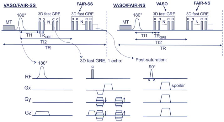
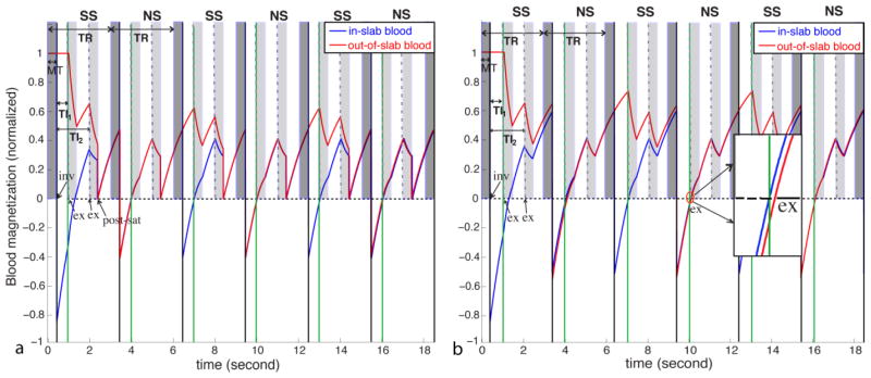
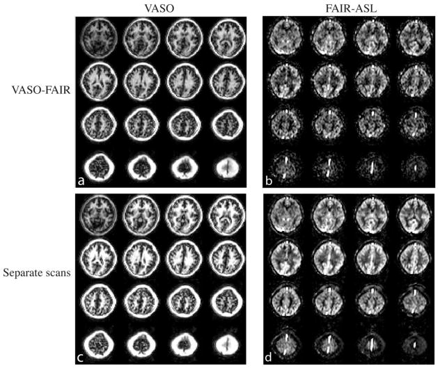
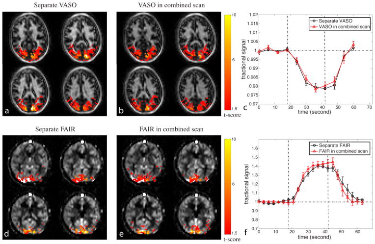
In addition to the BOLD scan, quantitative functional MRI studies require measurement of both cerebral blood volume (CBV) and flow (CBF) dynamics. The ability to detect CBV and CBF responses in a single additional scan would shorten the total scan time and reduce temporal variations. Several approaches for simultaneous CBV and CBF measurement during functional MRI experiments have been proposed in two-dimensional (2D) mode covering one to three slices in one repetition time (TR). Here, we extended the principles from previous work and present a three-dimensional (3D) whole-brain MRI approach that combines the vascular-space-occupancy (VASO) and flow-sensitive alternating inversion recovery (FAIR) arterial spin labeling (ASL) techniques, allowing the measurement of CBV and CBF dynamics, respectively, in a single scan. 3D acquisitions are complicated for such a scan combination as the time to null blood signal during a steady state needs to be known. We estimated this using Bloch simulations and demonstrate that the resulting 3D acquisition can detect activation patterns and relative signal changes of quality comparable to that of the original separate scans. The same was found for temporal signal-to-noise ratio (SNR) and contrast-to-noise ratio (CNR). This approach provides improved acquisition efficiency when both CBV and CBF responses need to be monitored during a functional task.
Keywords: ASL; Arterial spin labeling; CBF; CBV; FAIR; Flow-sensitive alternating inversion recovery; Three-dimensional; VASO; Vascular-space-occupancy; Whole-brain; fMRI.
Copyright © 2014 Elsevier Inc. All rights reserved.
Figures
Similar articles
-
A three-dimensional single-scan approach for the measurement of changes in cerebral blood volume, blood flow, and blood oxygenation-weighted signals during functional stimulation.Neuroimage. 2017 Feb 15;147:976-984. doi: 10.1016/j.neuroimage.2016.12.082. Epub 2016 Dec 29. Neuroimage. 2017. PMID: 28041979
-
Comparison of 3T and 7T ASL techniques for concurrent functional perfusion and BOLD studies.Neuroimage. 2017 Aug 1;156:363-376. doi: 10.1016/j.neuroimage.2017.05.038. Epub 2017 May 19. Neuroimage. 2017. PMID: 28528845
-
Theoretical and experimental investigation of the VASO contrast mechanism.Magn Reson Med. 2006 Dec;56(6):1261-73. doi: 10.1002/mrm.21072. Magn Reson Med. 2006. PMID: 17075857
-
Non-BOLD contrast for laminar fMRI in humans: CBF, CBV, and CMRO2.Neuroimage. 2019 Aug 15;197:742-760. doi: 10.1016/j.neuroimage.2017.07.041. Epub 2017 Jul 20. Neuroimage. 2019. PMID: 28736310 Review.
-
Noninvasive functional imaging of cerebral blood volume with vascular-space-occupancy (VASO) MRI.NMR Biomed. 2013 Aug;26(8):932-48. doi: 10.1002/nbm.2905. Epub 2013 Jan 28. NMR Biomed. 2013. PMID: 23355392 Free PMC article. Review.
Cited by
-
Techniques for blood volume fMRI with VASO: From low-resolution mapping towards sub-millimeter layer-dependent applications.Neuroimage. 2018 Jan 1;164:131-143. doi: 10.1016/j.neuroimage.2016.11.039. Epub 2016 Nov 18. Neuroimage. 2018. PMID: 27867088 Free PMC article.
-
Impaired response of cerebral oxygen metabolism to visual stimulation in Huntington's disease.J Cereb Blood Flow Metab. 2021 May;41(5):1119-1130. doi: 10.1177/0271678X20949286. Epub 2020 Aug 17. J Cereb Blood Flow Metab. 2021. PMID: 32807001 Free PMC article.
References
-
- Alsop DC, Detre JA. Reduced transit-time sensitivity in noninvasive magnetic resonance imaging of human cerebral blood flow. J Cereb Blood Flow Metab. 1996;16:1236–1249. - PubMed
-
- Alsop DC, Detre JA, Golay X, Gunther M, Hendrikse J, Hernandez-Garcia L, Lu H, Macintosh BJ, Parkes LM, Smits M, van Osch MJ, Wang DJ, Wong EC, Zaharchuk G. Recommended implementation of arterial spin-labeled perfusion MRI for clinical applications: A consensus of the ISMRM perfusion study group and the European consortium for ASL in dementia. Magn Reson Med. 2014 doi: 10.1002/mrm.25197.. - DOI - PMC - PubMed
-
- Balaban RS, Chesnick S, Hedges K, Samaha F, Heineman FW. Magnetization transfer contrast in MR imaging of the heart. Radiology. 1991;180:671–675. - PubMed
-
- Blockley NP, Francis ST, Gowland PA. Perturbation of the BOLD response by a contrast agent and interpretation through a modified balloon model. Neuroimage. 2009;48:84–93. - PubMed
Publication types
MeSH terms
Substances
Grants and funding
LinkOut - more resources
Full Text Sources
Other Literature Sources
Medical
