. 2014 Sep 4;95(3):285-93.
doi: 10.1016/j.ajhg.2014.07.012. Epub 2014 Aug 21.

Neu-Laxova syndrome is a heterogeneous metabolic disorder caused by defects in enzymes of the L-serine biosynthesis pathway

Affiliations

Neu-Laxova syndrome is a heterogeneous metabolic disorder caused by defects in enzymes of the L-serine biosynthesis pathway

Rocio Acuna-Hidalgo et al. Am J Hum Genet. .

Abstract

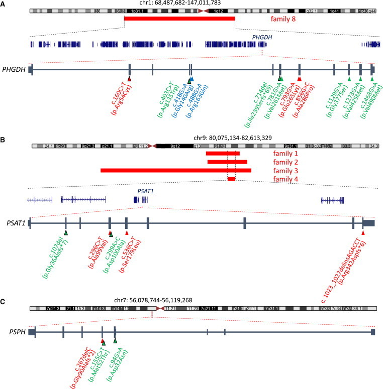
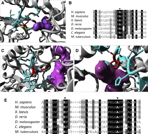
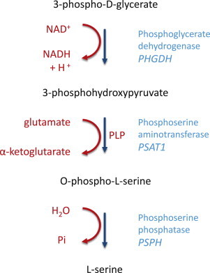
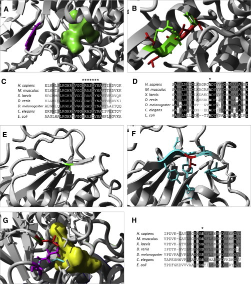
Neu-Laxova syndrome (NLS) is a rare autosomal-recessive disorder characterized by a recognizable pattern of severe malformations leading to prenatal or early postnatal lethality. Homozygous mutations in PHGDH, a gene involved in the first and limiting step in L-serine biosynthesis, were recently identified as the cause of the disease in three families. By studying a cohort of 12 unrelated families affected by NLS, we provide evidence that NLS is genetically heterogeneous and can be caused by mutations in all three genes encoding enzymes of the L-serine biosynthesis pathway. Consistent with recently reported findings, we could identify PHGDH missense mutations in three unrelated families of our cohort. Furthermore, we mapped an overlapping homozygous chromosome 9 region containing PSAT1 in four consanguineous families. This gene encodes phosphoserine aminotransferase, the enzyme for the second step in L-serine biosynthesis. We identified six families with three different missense and frameshift PSAT1 mutations fully segregating with the disease. In another family, we discovered a homozygous frameshift mutation in PSPH, the gene encoding phosphoserine phosphatase, which catalyzes the last step of L-serine biosynthesis. Interestingly, all three identified genes have been previously implicated in serine-deficiency disorders, characterized by variable neurological manifestations. Our findings expand our understanding of NLS as a disorder of the L-serine biosynthesis pathway and suggest that NLS represents the severe end of serine-deficiency disorders, demonstrating that certain complex syndromes characterized by early lethality could indeed be the extreme end of the phenotypic spectrum of already known disorders.


Figures

Figure 1
Figure 1
Identification of Mutations in PHGDH, PSAT1, and PSPH in Individuals with NLS by Homozygosity Mapping and Exome and Sanger Sequencing (A) In one family (family 8), a large homozygous region (68.4–147.0 Mb) on chromosome 1 was identified. This region overlaps the gene PHGDH, in which a mutation has recently been identified to cause NLS. The mutations found in families 7–9 are depicted at the cDNA level (red triangles) with their protein consequences below. PHGDH mutations were identified in a total of three individuals with NLS. These are compared to PHGDH mutations previously identified in individuals with serine-deficiency syndrome (MIM 614023; green triangles) and NLS (blue triangles). (B) Schematic overview of homozygous regions mapped to chromosome 9 in affected individuals from families 1–4. Overlapping homozygous regions (red bars) were identified in four families (family 1 by WES, families 2–4 by microarray analysis); the smallest region of overlap (80.1–82.6 Mb) mapped to chromosome 9. This region contains five protein-coding RefSeq genes (shown in blue), one of which is PSAT1. The mutations found in families 1–6 are depicted at the cDNA level (red triangles) with their protein consequences below. PSAT1 mutations were identified in a total of six individuals with NLS. These are compared to PSAT1 mutations previously identified in an individual with serine-deficiency syndrome (green triangles). (C) In one family (family 10), we identified a homozygous PSPH frameshift mutation (56.0–56.1 Mb) located on chromosome 7. The mutations are depicted at the cDNA level (red triangle) with their protein consequences below. A PSPH mutation has been previously reported in a child with serine-deficiency syndrome and Williams-Beuren syndrome (green triangle). Triangles outlined in black depict heterozygous mutations, whereas triangles with no outline depict homozygous mutations.
Figure 2
Figure 2
Modeling of PHGDH Substitutions (A, C, and D) 3D models of human PHGDH show in red the substitutions p.Arg54Cys (A), p.Glu265Lys (C), and p.Ala286Pro (D). All three substitutions lead to clashes with the side chains of neighboring residues (cyan) in close proximity to the binding site of phosphoglycerate and NAD+ (shown in purple). (B and E) Alignment of PHGDH sequence from multiple organisms shows high conservation of residues Arg54 (B, marked by an asterisk) and Glu265 and Ala286 (E, marked by the left and right asterisks, respectively). Protein sequences for PHGDH orthologs in Homo sapiens (UniProt ID O43175), Mus musculus (UniProt ID Q61753), Xenopus laevis (RefSeq NP_001015929.1), Danio rerio (RefSeq NP_955871.1), Drosophila melanogaster (RefSeq NP_609496.1), Caenorhabditis elegans (RefSeq NP_496868.1), and Mycobacterium tuberculosis (UniProt ID P9WNX3) were obtained from UniProt and Entrez. The 3D structure of wild-type human PHGDH (Protein Data Bank [PDB] ID 2G76) was obtained from the PDB, and substitutions were modeled with the FoldX plugin for YASARA.
Figure 3
Figure 3
L-Serine Biosynthesis Pathway De novo biosynthesis of serine is crucial to provide the organism with sufficient levels of this amino acid. The first and limiting step of this pathway is the conversion of 3-phosphoglycerate to 3-phosphohydroxypiruvate by PHGDH (encoded by PHGDH). This is followed by the conversion of 3-phosphohydroxypiruvate to O-phosphoserine by PSAT (encoded by PSAT1). This reaction is accompanied by the transformation of glutamate to α-ketoglutarate and requires the presence of pyridoxal phosphate (PLP). The final step in serine biosynthesis is catalyzed by PSPH (encoded by PSPH), which gives rise to L-serine, the final product of this pathway.
Figure 4
Figure 4
Modeling of PSAT1 Substitutions (A) A 3D model of PSAT from Escherichia coli shows glutamate (molecule in green) localizing closely to the beta sheet where Arg335 (corresponding to human Arg342 and highlighted in purple) is located. (B) Modeling of multiple residue substitutions resulting from frameshift p.Arg342fs6, in which the side chain of the wild-type residues (in green) overlaps the side chain of the altered residues (in red). (C) Alignment of PSAT sequence from multiple organisms shows the high conservation of the region surrounding Arg342. The seven altered residues resulting from the frameshift are marked by an asterisk. (D) Alignment of PSAT sequence shows high conservation of Ala99 (marked by an asterisk). (E and F) 3D model of human PSAT substitution p.Ala99Val. Residue Ala99 of PSAT is located in a beta sheet (E, in green). Substitution p.Ala99Val (F, in red) is predicted to disrupt the beta sheet by creating clashes between the large side chains of neighboring residues (shown in cyan). (G) A 3D model of PSAT from E. coli (in white) shows its binding to PLP (molecule in yellow) through the side chain of Lys198 (corresponding to human Lys200, in cyan). When residue Ser177 (corresponding to human Ser179, shown in green) is changed to a leucine (visible in red), the side chain of Lys198 (purple) is displaced and interferes with the binding of PLP. (H) Alignment of PSAT sequences from multiple organisms shows high conservation of residue S179. Protein sequences for PSAT1 orthologs in Homo sapiens (UniProt ID Q9Y617), Mus musculus (UniProt ID Q99K85), Xenopus laevis (RefSeq NP_001016582.1), Danio rerio (RefSeq NP_956113.1), Drosophila melanogaster (UniProt ID Q9VAN0), Caenorhabditis elegans (UniProt ID P91856), and Escherichia coli (UniProt ID Q8XEA7) were obtained from Uniprot and Entrez. 3D models for human PSAT (PDB ID 3E77) and E. coli serC (PDB ID 1BJO11) were obtained from the PDB.

References

    1. Neu R.L., Kajii T., Gardner L.I., Nagyfy S.F. A lethal syndrome of microcephaly with multiple congenital anomalies in three siblings. Pediatrics. 1971;47:610–612. - PubMed
    1. Laxova R., Ohara P.T., Timothy J.A. A further example of a lethal autosomal recessive condition in sibs. J. Ment. Defic. Res. 1972;16:139–143. - PubMed
    1. Manning M.A., Cunniff C.M., Colby C.E., El-Sayed Y.Y., Hoyme H.E. Neu-Laxova syndrome: detailed prenatal diagnostic and post-mortem findings and literature review. Am. J. Med. Genet. A. 2004;125A:240–249. - PubMed
    1. Shaheen R., Rahbeeni Z., Alhashem A., Faqeih E., Zhao Q., Xiong Y., Almoisheer A., Al-Qattan S.M., Almadani H.A., Al-Onazi N. Neu-Laxova syndrome, an inborn error of serine metabolism, is caused by mutations in PHGDH. Am. J. Hum. Genet. 2014;94:898–904. - PMC - PubMed
    1. Tabatabaie L., de Koning T.J., Geboers A.J., van den Berg I.E., Berger R., Klomp L.W. Novel mutations in 3-phosphoglycerate dehydrogenase (PHGDH) are distributed throughout the protein and result in altered enzyme kinetics. Hum. Mutat. 2009;30:749–756. - PubMed

Publication types

MeSH terms

Supplementary concepts