Review
. 2014 Oct:63:235-48.
doi: 10.1016/j.neuropsychologia.2014.08.013. Epub 2014 Aug 23.

The amygdala as a hub in brain networks that support social life

Affiliations
Review

The amygdala as a hub in brain networks that support social life

Kevin C Bickart et al. Neuropsychologia. 2014 Oct.

Abstract

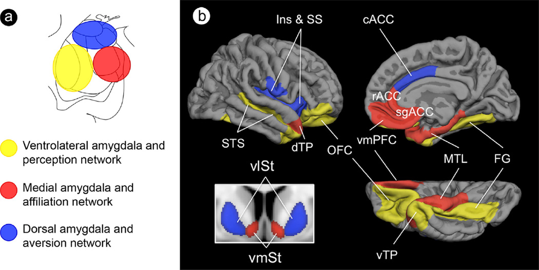
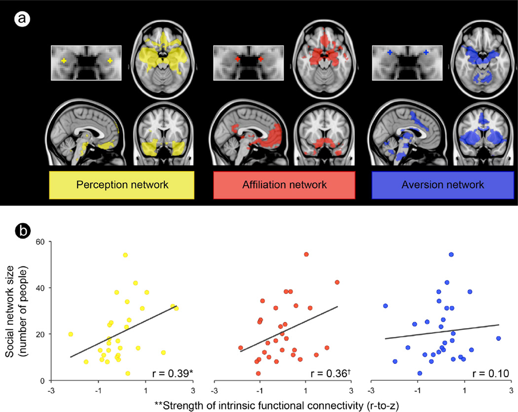
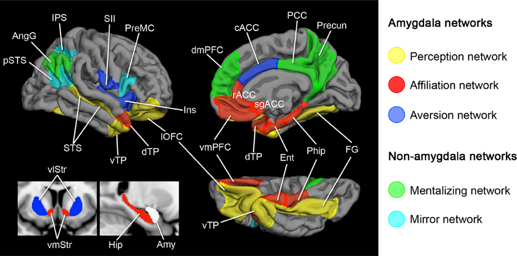
A growing body of evidence suggests that the amygdala is central to handling the demands of complex social life in primates. In this paper, we synthesize extant anatomical and functional data from rodents, monkeys, and humans to describe the topography of three partially distinct large-scale brain networks anchored in the amygdala that each support unique functions for effectively managing social interactions and maintaining social relationships. These findings provide a powerful componential framework for parsing social behavior into partially distinct neural underpinnings that differ among healthy people and disintegrate or fail to develop in neuropsychiatric populations marked by social impairment, such as autism, antisocial personality disorder, and frontotemporal dementia.

Keywords: Amygdala; Networks; Social brain; Social life; Social network.


Figures

Fig. 1
Fig. 1
Topographic schematic of amygdala subregions and their affiliated large-scale networks subserving social cognition. A schematic of (a) the amygdala subregions in coronal view that are anchors for (b) three large-scale networks subserving processes important for social cognition. Abbreviations: Ins, insula; SS, somatosensory operculum; STS, superior temporal sulcus; dTP, dorsal temporal pole; OFC, orbitofrontal cortex; cACC, caudal anterior cingulate cortex; rACC, rostral anterior cingulate cortex; sgACC, subgenual anterior cingulate cortex; vmPFC, ventromedial prefrontal cortex; MTL, medial temporal lobe; FG, fusiform gyrus; vTP, ventral temporal pole; vlSt, ventrolateral striatum; vmSt, ventromedial striatum. (For interpretation of the references to color in this figure, the reader is referred to the web version of this article.)
Fig. 2
Fig. 2
A larger social network is predicted by stronger connectivity between amygdala subregions and corticolimbic regions important for perception and affiliative behavior. (a) Each amygdala subregion and its intrinsic connectivity network were independently defined in the discovery sample (shown here) and then used in the test sample to compute the connectivity strength between each amygdala subregion and the rest of the network. (b) Scatter plots show that social network size (y-axis) is predicted by the strength of connectivity between two of the three amygdala subregions and their respective networks (x-axis), over and above amygdala volume, in the test sample. *p<0.05; p=0.06; **The x-axis displays the residual variance in the strength of the resting-state connectivity measure (Fisher’s r-to-z) after partialling out its shared variance with amygdala volume.
Fig. 3
Fig. 3
Exploratory analyses revealed that the connectivity between amygdala and specific regions within the perception and affiliation networks are the best predictors of social network size. (a) Brain images show location of voxels within the medial and ventrolateral amygdala’s intrinsic connectivity networks (defined in the discovery sample) that correlated with social network size at p <0.01 in the test sample, uncorrected with a cluster size constraint of 10 voxels. Color bars indicate the p-values (10−2–10−4) of correlated voxels, which are overlaid on slices of a T1 MNI152 0.5 mm template brain in radiologic convention. (b) Scatter plots show the effects for peak voxels with the strength of intrinsic connectivity on the y-axis and social network size on the x-axis. **p< 0.001.
Fig. 4
Fig. 4
A schematic of five large-scale brain networks subserving processes important for social behavior. Here, we show three networks that are anchored in the amygdala (amygdala-based networks) and two that are not (control networks). The amygdala is displayed in white indicating that it is the hub of the three amygdala-based networks. List of ROIs with abbreviations–Perception network: lOFC, lateral orbitofrontal cortex; vTP, ventrolateral temporal pole; FG, fusiform gyrus; STS, superior temporal sulcus. Affiliation network: dTP, dorsomedial temporal pole; rACC, rostral anterior cingulate cortex; sgACC, subgenual anterior cingulate cortex; vmPFC, ventromedial prefrontal cortex; Ent, entorhinal cortex; PHip, parahippocampal cortex; vmSt, ventromedial striatum. Aversion network: cACC, caudal anterior cingulate cortex; Ins, insula; SII, somatosensory operculum; vlSt, ventrolateral striatum. Mentalizing network: dmPFC, dorsomedial prefrontal cortex; PCC, posterior cingulate cortex; Precun, precuneus; AngG, angular gyrus (temporoparietal junction). Mirror network: pSTS, posterior superior temporal sulcus; IPS, intraparietal sulcus; PreMC, premotor cortex.

References

    1. Adolphs R. Social cognition and the human brain. Trends in Cognitive Sciences. 1999;3:469–479. - PubMed
    1. Adolphs R. The neurobiology of social cognition. Current Opinion in Neurobiology. 2001;11:231–239. - PubMed
    1. Adolphs R. The social brain: neural basis of social knowledge. Annual Review of Psychology. 2009;60:693–716. - PMC - PubMed
    1. Adolphs R, Tranel D, Damasio AR. The human amygdala in social judgment. Nature. 1998;393:470–474. - PubMed
    1. Adolphs R, Tranel D, Damasio H, Damasio AR. Fear and the human amygdala. Journal of Neuroscience. 1995;15:5879–5891. - PMC - PubMed

Publication types