. 2008 Dec 1;4(4):439-449.
doi: 10.1166/jbn.2008.007.

Development of Receptor Targeted Magnetic Iron Oxide Nanoparticles for Efficient Drug Delivery and Tumor Imaging

Affiliations

Development of Receptor Targeted Magnetic Iron Oxide Nanoparticles for Efficient Drug Delivery and Tumor Imaging

Lily Yang et al. J Biomed Nanotechnol. .

Abstract

The development of multifunctional nanoparticles that have dual capabilities of tumor imaging and delivering therapeutic agents into tumor cells holds great promises for novel approaches for tumor imaging and therapy. We have engineered urokinase plasminogen activator receptor (uPAR) targeted biodegradable nanoparticles using a size uniform and amphiphilic polymer-coated magnetic iron oxide (IO) nanoparticle conjugated with the amino-terminal fragment (ATF) of urokinase plasminogen activator (uPA), which is a high affinity natural ligand for uPAR. We further developed methods to encapsulate hydrophobic chemotherapeutic drugs into the polymer layer on the IO nanoparticles, making these targeted magnetic resonance imaging (MRI) sensitive nanoparticles drug delivery vehicles. Using a fluorescent drug doxorubicin (Dox) as a model system, we showed that this hydrophobic drug can be efficiently encapsulated into the uPAR-targeted IO nanoparticles. This class of Dox-loaded nanoparticles has a compact size and is stable in pH 7.4 buffer. However, encapsulated Doxcan be released from the nanoparticles at pH 4.0 to 5.0 within 2 hrs. In comparison with the effect of equivalent dosage of free drug or non-targeted IO-Dox nanoparticles, uPAR-targeted IO-Dox nanoparticles deliver higher levels of Dox into breast cancer cells and produce a stronger inhibitory effect on tumor cell growth. Importantly, Dox-loaded IO nanoparticles maintain their T2 MRI contrast effect after being internalized into the tumor cells due to their significant susceptibility effect in the cells, indicating that this drug delivery nanoparticle has the potential to be used as targeted therapeutic imaging probes for monitoring the drug delivery using MRI.

Keywords: Breast Cancer; Doxorubicin; Drug Delivery Nanoparticle; Magnetic Iron Oxide Nanoparticles; Targeted Nanoparticle; uPAR.


Figures

Fig. 1
Fig. 1
Examination of the binding specificity of the recombinant ATF peptide and its ability to target IO drug-nanoparticles into breast cancer cells. (A) IO nanoparticles synthesized have a uniformed size of 10 nm as shown in transmission electron microscopy (TEM). (B) Examination of purified mouse ATF peptides by SDS-PAGE gel. ATF peptides appeared as monomers (17 KDa) or dimers (35 KDa). (C) Detection of the binding of mouse ATF peptides to mouse mammary tumor 4T1 cells. Red fluorescence is found on the surface of tumor cells after incubating ATF peptides followed by mouse anti-His tag antibody and Alexa-fluor 555 labeled anti-mouse antibody. However, red fluorescence is not detected in the cells incubated with anti-His tag and secondary antibodies without ATF-peptides. Blue: Hoechst 33324 background staining. (D) Mouse ATF conjugated-IO nanoparticles are able to bind to and be internalized by human breast cancer MDA-MB-231 cells. Prussian blue staining shows strong blue iron staining inside the cells incubated with ATF-IO nanoparticles for 2 hrs. Cells incubated with non-targeted IO only display a low level of non-specific iron staining. (E) Mouse mammary tumor 4T1 cells were incubated with 1 μM of free Dox or equivalent Dox dosages of IO-Dox and ATF-IO-Dox for 24 to 48 hrs. Cells were examined under a fluorescence microscope. After incubating with free Dox, most cells showed weak fluorescence signal with a small percentage of the cells having a higher level of Dox. Non-targeted IO-Dox treated cells have an increased amount of Dox in all tumor cells, suggesting that non-specific uptake of IO nanoparticles at a higher concentration of IO nanoparticles and after longer incubation time can also deliver the drug into the cells. However, tumor cells treated with uPAR targeted, ATF-IO-Dox nanoparticles have a significant higher level of Dox that is located in the cytoplasm of the cells at 24 hrs and then moved to cell nucleus at 48 hrs. Interestingly, Dox fluorescence at 48 hrs showed fragmented nuclear staining, which is an indication of apoptotic cell death induced by incorporating Dox into cellular DNA (far right picture).
Fig. 2
Fig. 2
Examination of pH sensitive release of Dox from non-targeted or ATF-targeted IO nanoparticles in vitro. Non-targeted and targeted-IO-Dox nanoparticles containing 400 to 600 pmol of Dox were placed in the buffer with pH 4, 5, 6 and 7 for 2 hrs at 37 °C. The amount of released Dox molecules was determined by measuring fluorescence intensity in solution and then calculated from the Dox standard curve. The total amount of Dox added to IO-Dox or ATF-IO-Dox nanoparticles is used as 100%. Result shown is the mean of three repeat experiments.
Fig. 3
Fig. 3
Detection of effect of drug delivery using ATF-IO-Dox on breast cancer cells in vitro. Cells cultured in 96 well plates were incubated with the medium containing 0.25 or 0.5 μM of free Dox, IO-Dox or ATF-IO-Dox nanoparticles as well as control IO and ATF-IO nanoparticles for 2 hrs. IO nanoparticles with 10 nm core size were used from this study. After removing the drug and nanoparticles, the cells were cultured for 48 hrs and then fixed with 4% formaldehyde in PBS buffer. (A and B) Examination of the viable cells and Dox fluorescence in 4T1 cells using phase contrast and fluorescence microscopy and cell proliferation assay. Treatment of the cells with 0.5 μM of free Dox and IO-Dox nanoparticles did not induce cell death in 4T1 cells. Dox fluorescence was absent or present in a very low level in those cells (A). However, a high level of Dox was detected in the tumor cells after 0.25 to 0.5 μM of ATF-IO-Dox treatment. Higher magnification fluorescence image (40× lens) shows the presence of Dox in the cell nucleus. Cell proliferation assay shows that treatment of 4T1 cells with ATF-IO-Dox nanoparticles at 0.5 μM of Dox concentration induces cell death in 75% of the cells, while the same drug concentration of free Dox or IO-DOX, or same IO concentration of IO or ATF-IO treated cells did not show any cytotoxic effects on the cells (B). In human breast cancer MDA-MB-231 cells, 0.5 μM of free Dox treatment did not induce cell death and Dox fluorescence was not detected in the cells (C and D). Our result showed non-specific uptake of IO-Dox by MDA-MB-231 cells also induced cell death. However, a higher level of Dox fluorescence and 40% more cell death were found in ATF-IO-Dox treated cells compared to IO-Dox treated cells (C and D). A slightly high percentage of MDA-MB-231 cells detected in ATF-IO treated group is due to the internalization of IO nanoparticles into cells resulting in an enhanced Crystal Violet staining. Results shown are the mean of four repeat samples. Similar results were obtained from three separate studies.
Fig. 4
Fig. 4
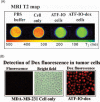
Examination of effect of drug encapsulation on the T 2 weighted MRI contrast produced by IO nanoparticles. Human breast cancer MDA-MB-231 cells were incubated with 20 pmol of ATF-IO or ATF-Dox-IO nanoparticles for 4 hrs.After washing with PBS, the cell pellets were examined using 4.7T MRI scanner. (A) MRI T2 relaxometry demonstrated that ATF-IO and ATF-IO-Dox labeled tumor cells have similar T 2 values of 112±10 and 118±10 ms, respectively. However, T 2 value in ATF-IO or ATF-IO-Dox labeled cells is three times lower compared to unlabeled cells (470±10 ms), suggesting that ATF-IO-Dox nanoparticle may be used as a targeted MRI contrast agent. T 2 values of each sample/well were calculated from multi-echo images by fitting the decay curve on a pixel-by-pixel basis using the non-linear mono-exponential algorithm of Mi =M0 *exp (–TEi/T 2). Echo time (TE) has a unit in million seconds as shown in the figure.Orange-red color in the scale bar indicates a high T 2 value and green color represents a low T 2 value.(B) ATF-IO-Dox nanoparticle-labeled MDA-MB-231 cells, but not un-labeled cells, showed strong Dox fluorescence inside the cells.
Scheme
Scheme
Schematic illustration of the procedure for production of receptor-targeted drug delivery nanoparticles.Magnetic iron oxide (IO) nanoparticles with core sizes of 5 or 10 nm were coated with amphiphilic copolymers modified with short PEG chains.Recombinant ATF peptides were conjugated to the carboxyl side groups mediated by EDAC.Dox molecules were then encapsulated onto the surface of the IO nanoparticles.

References

    1. Jemal A, Siegel R, Ward E, Hao Y, Xu J, Murray T, Thun MJ. Cancer statistics. CA Cancer J. Clin. 2008;58:71. - PubMed
    1. Nasongkla N, Bey E, Ren J, Ai H, Khemtong C, Guthi JS, Chin SF, Sherry AD, Boothman DA, Gao J. Multifunctional polymeric micelles as cancer-targeted, MRI-ultrasensitive drug delivery systems. Nano Lett. 2006;6:2427. - PubMed
    1. Kohler N, Sun C, Fichtenholtz A, Gunn J, Fang C, Zhang M. Methotrexate-immobilized poly(ethylene glycol) magnetic nanoparticles for MR imaging and drug delivery. Small. 2006;2:785. - PubMed
    1. Medarova Z, Pham W, Farrar C, Petkova V, Moore A. In vivo imaging of siRNA delivery and silencing in tumors. Nat. Med. 2007;13:372. - PubMed
    1. Chen W, Zhang J. Using nanoparticles to enable simultaneous radiation and photodynamic therapies for cancer treatment. J. Nanosci. Nanotechnol. 2006;6:1159. - PubMed

LinkOut - more resources