Review
. 2014 Aug 8:8:76.
doi: 10.3389/fnana.2014.00076. eCollection 2014.

Lipid dynamics at dendritic spines

Affiliations
Review

Lipid dynamics at dendritic spines

Carlos Gerardo Dotti et al. Front Neuroanat. .

Abstract

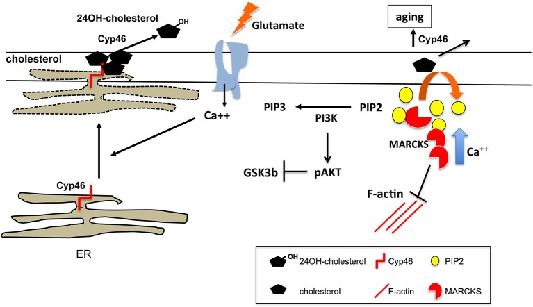
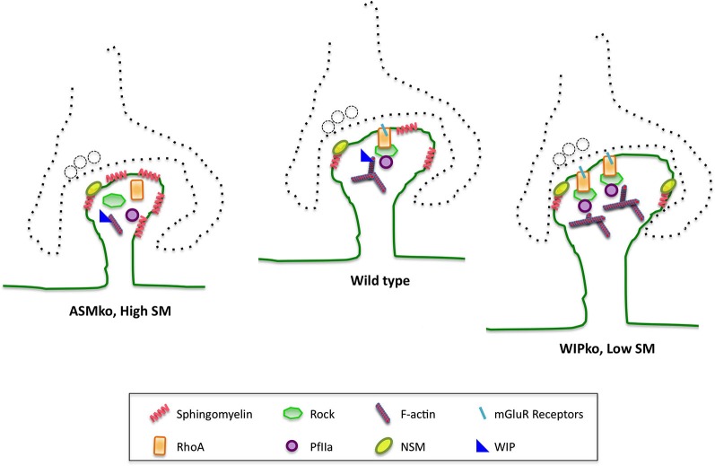
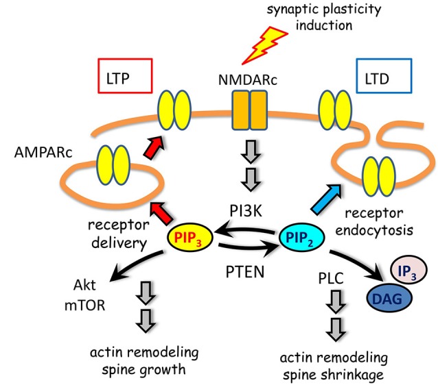
Dynamic changes in the structure and composition of the membrane protrusions forming dendritic spines underlie memory and learning processes. In recent years a great effort has been made to characterize in detail the protein machinery that controls spine plasticity. However, we know much less about the involvement of lipids, despite being major membrane components and structure determinants. Moreover, protein complexes that regulate spine plasticity depend on specific interactions with membrane lipids for proper function and accurate intracellular signaling. In this review we gather information available on the lipid composition at dendritic spine membranes and on its dynamics. We pay particular attention to the influence that spine lipid dynamism has on glutamate receptors, which are key regulators of synaptic plasticity.

Keywords: cholesterol; dendritic spines; glutamate receptors; phosphoinositides; sphingolipids; synaptic plasticity.


Figures

Figure 1
Figure 1
Cholesterol regulation at spines upon glutamate stimulation. Acute glutamate stimulatory conditions lead to loss of membrane cholesterol. The mechanism proposed involves glutamate induced rise in intracellular Ca++ leading to the approximation/apposition of ER membranes to the synaptic plasma membrane. This allows Cyp46A1, whose active site is in the lumenal side of the ER, to oxidize cholesterol present in the exoplasmic leaflet that is released as hydroxycholesterol. In the aging context, constitutive high intracellular calcium and irreversible cholesterol loss due to lifelong lasting synaptic activity leads to reduced membrane-associated MARCKS, which affects synaptic plasticity by several mechanisms: (1) impaired MARCKS-mediated actin dynamics; (2) reduced membrane clustering of PIP2; and (3) high PI3K activity resulting in reduced glutamate-mediated Akt dephosphorylation and GSK3β activation. The later contributes to the impaired AMPARc internalization and LTD in aged neurons.
Figure 2
Figure 2
SM and its catabolic enzymes in dendritic spine physiology and pathology. SM levels at the postsynaptic membrane modulate membrane binding and activation of the small GTPase RhoA, which in turn modulates F-actin content through its effectors ROCK and profillin 2A. This molecular mechanism controls dendritic spine size. The SM catabolic enzymes ASM and NSM are involved in this process. Hence, mice lacking ASM show abnormally high SM levels in their postsynaptic membranes that lower the amount of cell surface mGluRs. This impairs RhoA membrane binding and pathway activation, which diminish spine F-actin content and size. These anomalies can be corrected by NSM activation. Conversely, mice lacking the actin related protein WIP show constitutively active NSM leading to reduced SM levels, which enhance RhoA membrane binding and pathway activation resulting in bigger dendritic spines with higher F-actin content. Spine anomalies could explain the cognitive deficits observed in ASMko mice, which are a model for Niemann Pick disease type A, and those of individuals carrying mutations in the region encoding for WIP.
Figure 3
Figure 3
PIP metabolism in spine plasticity. Activation of NMDARc modulate AMPARc trafficking through spatially and timely controlled activity of PIPs and their metabolic enzymes. On one hand, PI3K association with AMPARc is required for receptor cell surface delivery during LTP. On the other hand, PTEN activity leading to PIP3 downregulation promotes migration of AMPARc from the postsynaptic density to the perisynaptic membrane. This depresses AMPARc synaptic responses by promoting receptor endocytosis during LTD. Moreover, signaling pathways initiated by PIP3 or PIP2, in which Akt/mTOR, DAG and IP3 are involved, contribute to actin remodeling and spine changes in size.

References

    1. Abe M., Makino A., Hullin-Matsuda F., Kamijo K., Ohno-Iwashita Y., Hanada K., et al. (2012). A role for sphingomyelin-rich lipid domains in the accumulation of phosphatidylinositol-4,5-bisphosphate to the cleavage furrow during cytokinesis. Mol. Cell. Biol. 32, 1396–1407 10.1128/MCB.06113-11 - DOI - PMC - PubMed
    1. Abulrob A., Tauskela J. S., Mealing G., Brunette E., Faid K., Stanimirovic D. (2005). Protection by cholesterol-extracting cyclodextrins: a role for N-methyl-D-aspartate receptor redistribution. J. Neurochem. 92, 1477–1486 10.1111/j.1471-4159.2005.03001.x - DOI - PubMed
    1. Allen J. A., Halverson-Tamboli R. A., Rasenick M. M. (2007). Lipid raft microdomains and neurotransmitter signalling. Nat. Rev. Neurosci. 8, 128–140 10.1038/nrn2059 - DOI - PubMed
    1. Arendt K. L., Royo M., Fernández-Monreal M., Knafo S., Petrok C. N., Martens J. R., et al. (2010). PIP3 controls synaptic function by maintaining AMPA receptor clustering at the postsynaptic membrane. Nat. Neurosci. 13, 36–44 10.1038/nn.2462 - DOI - PMC - PubMed
    1. Arroyo A. I., Camoletto P. G., Morando L., Sassoe-Pognetto M., Giustetto M., Van Veldhoven P. P., et al. (2014). Pharmacological reversion of sphingomyelin-induced dendritic spine anomalies in a Niemann pick disease type a mouse model. EMBO Mol. Med. 6, 398–413 10.1002/emmm.201302649 - DOI - PMC - PubMed