

Finite Element Modeling of CNS White Matter Kinematics: Use of a 3D RVE to Determine Material Properties
- PMID: 25152875
- PMCID: PMC4126384
- DOI: 10.3389/fbioe.2013.00019
Finite Element Modeling of CNS White Matter Kinematics: Use of a 3D RVE to Determine Material Properties
Abstract
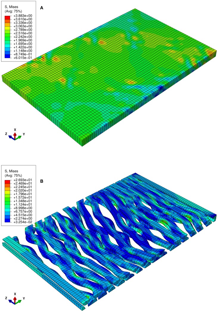
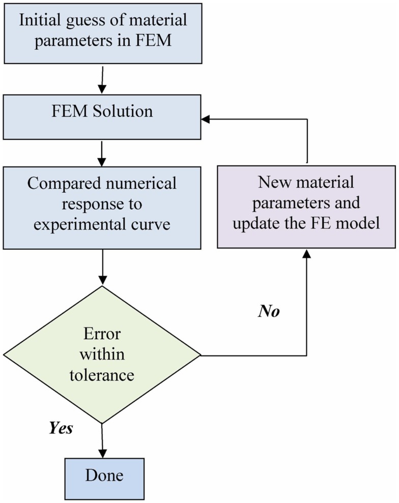
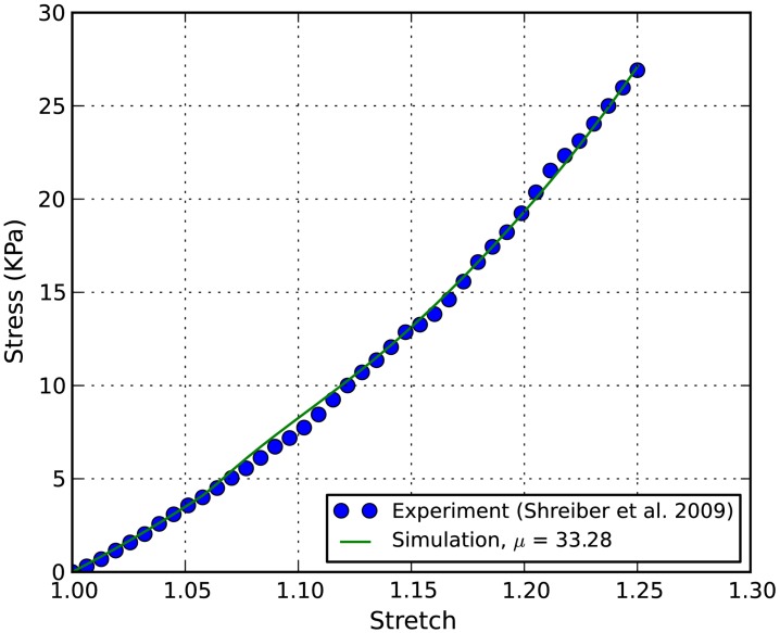
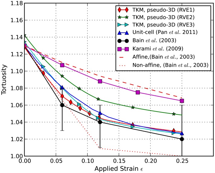
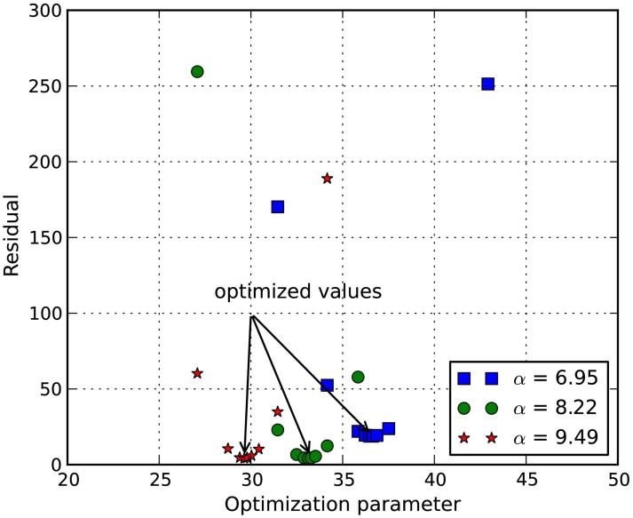
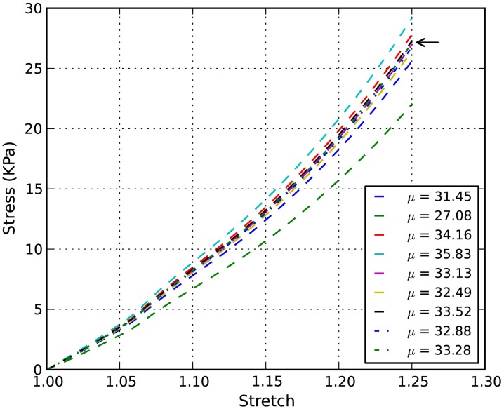
Axonal injury represents a critical target area for the prevention and treatment of traumatic brain and spinal cord injuries. Finite element (FE) models of the head and/or brain are often used to predict brain injury caused by external mechanical loadings, such as explosive waves and direct impact. The accuracy of these numerical models depends on correctly determining the material properties and on the precise depiction of the tissues' microstructure (microscopic level). Moreover, since the axonal microstructure for specific regions of the brain white matter is locally oriented, the stress, and strain fields are highly anisotropic and axon orientation dependent. Additionally, mechanical strain has been identified as the proximal cause of axonal injury, which further demonstrates the importance of this multi-scale relationship. In this study, our previously developed FE and kinematic axonal models are coupled and applied to a pseudo 3-dimensional representative volume element of central nervous system white matter to investigate the multi-scale mechanical behavior. An inverse FE procedure was developed to identify material parameters of spinal cord white matter by combining the results of uniaxial testing with FE modeling. A satisfactory balance between simulation and experiment was achieved via optimization by minimizing the squared error between the simulated and experimental force-stretch curve. The combination of experimental testing and FE analysis provides a useful analysis tool for soft biological tissues in general, and specifically enables evaluations of the axonal response to tissue-level loading and subsequent predictions of axonal damage.
Keywords: axonal injury; microstructural properties; multi-scale modeling; spinal cord injury; traumatic brain injury.
Figures
References
-
- Bain A. C., Billiar K. L., Shreiber D. I., McIntosh T. K., Meaney D. F. (1997). In vivo mechanical thresholds for traumatic axonal damage. Advisory Group for Aerospace Research and Development (AGARD) Meeting 199 (Mescalero, NM: ), 13 p.
LinkOut - more resources
Full Text Sources
Other Literature Sources