Review
. 2014 Aug 22;6(8):3363-76.
doi: 10.3390/v6083363.

Canine enteric coronaviruses: emerging viral pathogens with distinct recombinant spike proteins

Affiliations
Review

Canine enteric coronaviruses: emerging viral pathogens with distinct recombinant spike proteins

Beth N Licitra et al. Viruses. .

Abstract

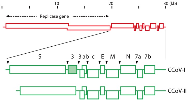
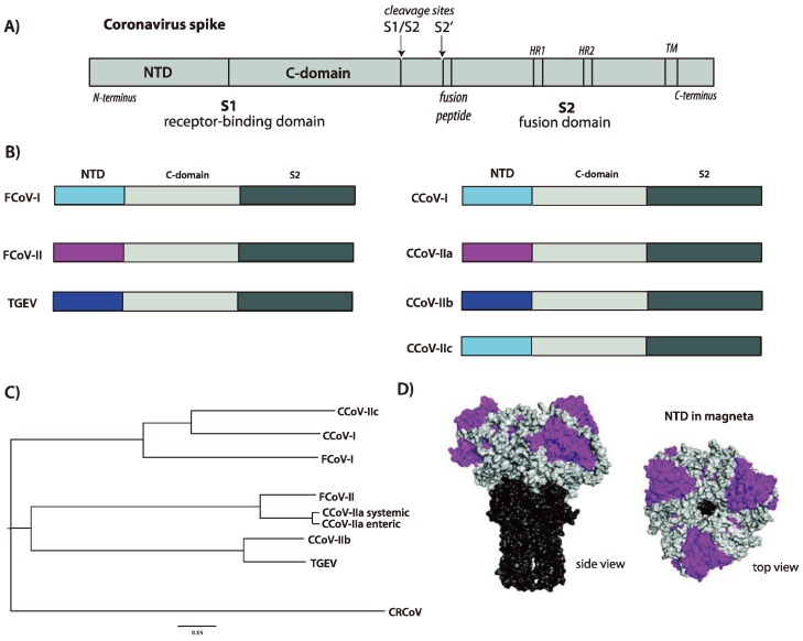
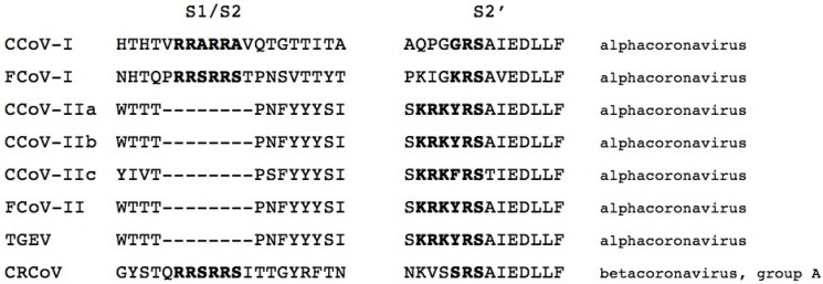
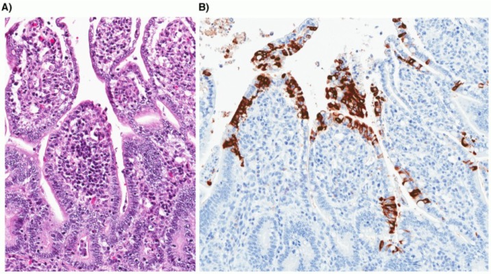
Canine enteric coronavirus (CCoV) is an alphacoronavirus infecting dogs that is closely related to enteric coronaviruses of cats and pigs. While CCoV has traditionally caused mild gastro-intestinal clinical signs, there are increasing reports of lethal CCoV infections in dogs, with evidence of both gastrointestinal and systemic viral dissemination. Consequently, CCoV is now considered to be an emerging infectious disease of dogs. In addition to the two known serotypes of CCoV, novel recombinant variants of CCoV have been found containing spike protein N-terminal domains (NTDs) that are closely related to those of feline and porcine strains. The increase in disease severity in dogs and the emergence of novel CCoVs can be attributed to the high level of recombination within the spike gene that can occur during infection by more than one CCoV type in the same host.


Figures

Figure 1
Figure 1
Image representation of the genomes of CCoV-1 and CCoV-II. Adapted from Lorusso et al. [22].
Figure 2
Figure 2
(A) Image representation of a coronavirus spike protein. Key features are identified, including the N-terminal domain (NTD) and C-domain within the S1 receptor-binding domain, and the fusion peptide, two heptad repeats (HR1 and HR2) and transmembrane domain (TM) in the S2 fusion domain. The two cleavage sites for protease activation (S1/S2 and S2’) are shown; (B) Image representation of the different domains present within the spike proteins of alpha-coronaviruses of cats (FCoV), dogs (CCoV) and pigs (TGEV). The different NTDs are color-coded to indicate homology and proposed recombination events across the species, with the remainder of S1 depicted in light gray and S2 in dark gray; (C) Phylogenetic tree of the NTDs present within the spike proteins of representative coronaviruses of cats (FCoV), dogs (CCoV) and pigs (TGEV) (The following virus sequences were used for alignment. NTD = aa 1-279 (based on CCoV-A76). FCoV-I = Black (BAC05493.1); FCoV-II = WSU-79-1146 (AGZ84516.1); CCoV-IIa = CCoV CB/05 (AAZ91437.1); CCoV-IIb = CCoV 341/05 (ACJ63231.1); CCoV-I = CCoV Elmo/02 (AAP72149.1); CCoV-IIc = CCoV-A76 (AEQ61968.1); CRCoV = CRCoV4182 (ABG78748.1); TGEV (CAB91145.1)). The tree was created using ClustalX [45] and FigTree software [46]; (D) Structural model of a coronavirus spike protein based on PDB file 1T7G [47], in surface rendering. The NTD is colored magenta, the remainder of S1 is light gray and the S2 domain is colored dark gray. Two views of the trimeric spike are shown, a side view and a top view.
Figure 3
Figure 3
Multiple sequence alignment of the spike proteins of representative coronaviruses of cats (FCoV) dogs (CCoV and CRCoV) and pigs (TGEV) (The following virus sequences were used for alignment. FCoV-I = RM (ACT10854); FCoV-II = WSU-79-1683 (AFH58021.1); CCoV-IIa = CCoV CB/05 (AAZ91437.1); CCoV-IIb = CCoV 341/05 (ACJ63231.1); CCoV-I = CCoV Elmo/02 (AAP72149.1); CCoV-IIc = CCoV-A76 (AEQ61968.1); CRCoV = CRCoV4182 (ABG78748.1); TGEV (CAB91145.1).), in the region of the two activation sites (S1/S2 and S2’). The alignment was created using ClustalX [45]. The expected cleavage motif is in bold.
Figure 4
Figure 4
Photomicrographs of small intestine taken from a two-week-old puppy with typical lesions of CCoV infection. (A) Severe atrophy of intestinal villi with attenuated low cuboidal to squamous enterocytes (hematoxylin and eosin stain; 20× original magnification). (B) Coronavirus antigen is present within the cytoplasm of infected villous enterocytes (immunohistochemistry; 20× original magnification).

References

    1. Binn L., Lazar E.C., Keenan K.P., Huxsoll D.L., Marchwicki R.H., Strano A.J. Recovery and characterization of a coronavirus from military dogs with diarrhea. Proc. Annu. Meet. U. S. Anim. Health Assoc. 1974;78:359–366. - PubMed
    1. Keenan K.P., Jervis H.R., Marchwicki R.H., Binn L.N. Intestinal infection of neonatal dogs with canine coronavirus 1-71: Studies by virologic, histologic, histochemical, and immunofluorescent techniques. Am. J. Vet. Res. 1976;37:247–256. - PubMed
    1. Perlman S., Gallagher T., Snijder E.J. Nidoviruses. ASM Press; Washington, DC, USA: 2008.
    1. Lai M.M.C., Holmes K.V. Coronaviridae: The Viruses and Their Replication. In: Knipe D.M., Howely P.M., editors. Fields Virology. Lippincott Wilkins and Williams; Philadelphia, PA, USA: 2001.
    1. Hogue B.G., Machamer C.E. Coronavirus Structural Proteins and Virus Assembly. In: Perlman S., Gallagher T., Snijder E.J., editors. Nidoviruses. ASM Press; Washington, DC, USA: 2008. pp. 179–200.

Publication types

MeSH terms

Substances