. 2014 Aug 15;5(15):6038-48.
doi: 10.18632/oncotarget.2153.

PTK6/BRK is expressed in the normal mammary gland and activated at the plasma membrane in breast tumors

Affiliations

PTK6/BRK is expressed in the normal mammary gland and activated at the plasma membrane in breast tumors

Maoyu Peng et al. Oncotarget. .

Abstract

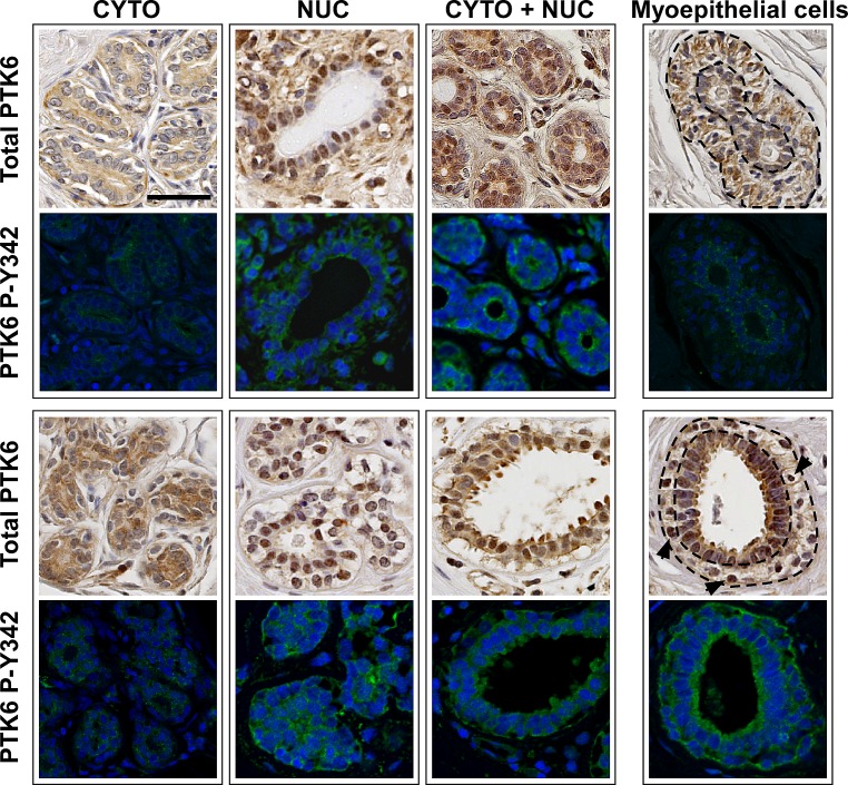
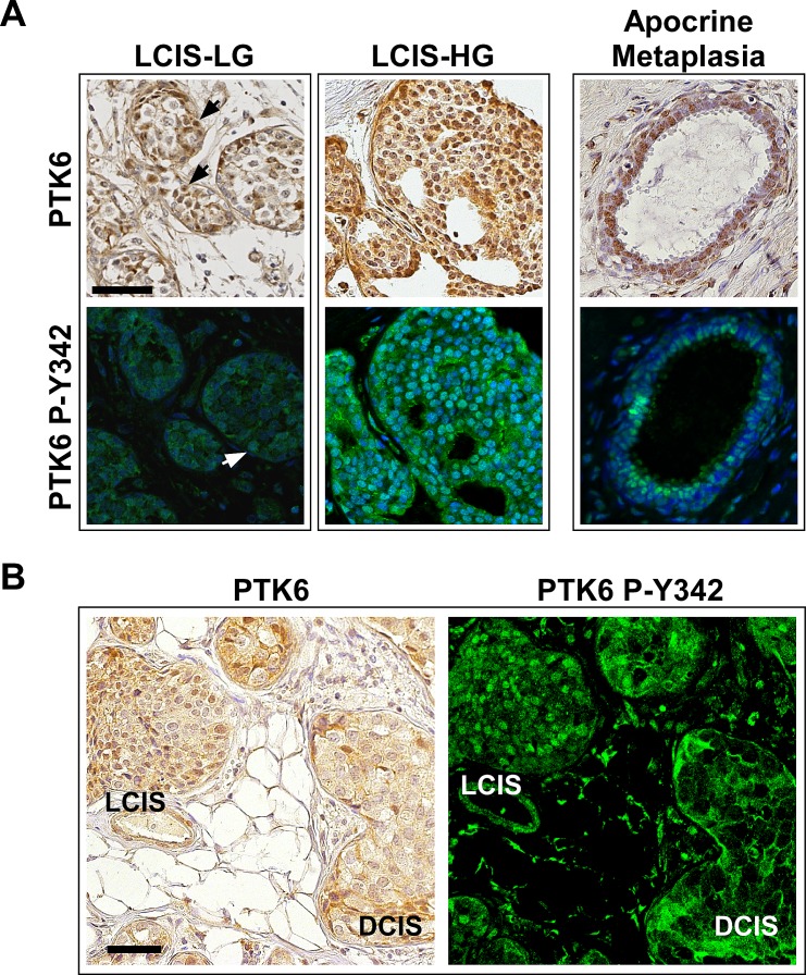
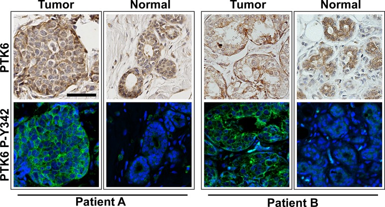
Protein Tyrosine kinase 6 (PTK6/BRK) is overexpressed in the majority of human breast tumors and breast tumor cell lines. It is also expressed in normal epithelial linings of the gastrointestinal tract, skin, and prostate. To date, expression of PTK6 has not been extensively examined in the normal human mammary gland. We detected PTK6 mRNA and protein expression in the immortalized normal MCF-10A human mammary gland epithelial cell line, and examined PTK6 expression and activation in a normal human breast tissue microarray, as well as in human breast tumors. Phosphorylation of tyrosine residue 342 in the PTK6 activation loop corresponds with its activation. Similar to findings in the prostate, we detect nuclear and cytoplasmic PTK6 in normal mammary gland epithelial cells, but no phosphorylation of tyrosine residue 342. However, in human breast tumors, striking PTK6 expression and phosphorylation of tyrosine 342 is observed at the plasma membrane. PTK6 is expressed in the normal human mammary gland, but does not appear to be active and may have kinase-independent functions that are distinct from its cancer promoting activities at the membrane. Understanding consequences of PTK6 activation at the plasma membrane may have implications for developing novel targeted therapies against this kinase.


Conflict of interest statement

Disclosure of Potential Conflicts of Interest:

The authors have no conflicts of interest to report. This is an original manuscript; data have not been published elsewhere

Figures

Figure 1
Figure 1. Controls were performed to confirm the specificity of PTK6 antibody for detection of PTK6 in nontransformed cells and tissues
(A) Total cell lysates from several human breast cell lines were probed with a 1:2000 dilution of PTK6 C-18 antibody (Santa Cruz), and the entire membrane is shown. A specific 50 kDa band corresponding to PTK6 is detected in all samples. (B) Specificity of the PTK6 C-18 antibody for immunohistochemistry was confirmed using serial sections of normal human mammary gland and PTK6 C-18 antibody plus and minus pre-incubation with the PTK6 immunogenic peptide. Blocking with PTK6 peptide eliminates detection of the cytoplasmic and nuclear staining obtained with the C-18 antibody. (C) P-Y342 PTK6 was detected only in human breast tumors. Normal mammary gland and tumors were stained with PTK6 P-Y342 antibody or rabbit IgG as a control. The same gain time for FITC was used when taking both PTK6 and IgG pictures (scale bar = 50 μm).
Figure 2
Figure 2. PTK6 is expressed in normal human mammary gland but it is not phosphorylated at tyrosine residue 342
Immunohistochemistry and immunofluorescence assays were performed on serial sections of human mammary gland tissue microarray. The TMA cores contained normal mammary glands obtained from breast reduction or benign biopsy. Antibodies against total human PTK6 and P-Y342 PTK6 were used to detect endogenous PTK6. Weak to medium levels of PTK6 were detected in luminal cells of most cores, and expression was cytoplasmic, nuclear or in a mixed pattern. Weak PTK6 expression was also detected in myoepithelial cells, and its subcellular localization varied, with it being predominately cytoplasmic (upper right corner, outlined by dashed lines) and in a few cases in the nuclei of vacuolated myoepithelial cells (arrows, lower right corner, outlined by dashed lines). However, active PTK6 P-Y342 (FITC green) was not detected in the normal mammary glands. Signal shown is equivalent to the IgG signal in Figure 1 (scale bar = 50 μm).
Figure 3
Figure 3. Active P-Y342 PTK6 is localized at the plasma membrane in high-grade ductal carcinomas
Immunohistochemistry and immunofluorescence assays were performed on breast tumor TMA slides using the same conditions as normal breast TMA. Medium to strong levels of total PTK6 protein immunoreactivity were detected in breast tumor tissues (IDC: invasive ductal carcinoma. DCIS: ductal carcinoma in situ. LG: low-grade. HG: high-grade). Total PTK6 and P-Y342 PTK6 were shown in boxed pairs, with each box representing a different patient, and two patients are shown for each tumor type (upper and lower panels) with the exception of the DCIS-HG where two different cores from the same patient are shown. Although some nuclear staining of total PTK6 can be found in the low-grade carcinoma (arrows), it was absent from most of the high-grade carcinomas and PTK6 appears to be in the cytoplasm and at the membrane. Similar expression patterns are observed in ductal carcinoma in situ. In the low-grade ductal carcinomas, the P-Y342 signal is low, but in the high-grade invasive ductal carcinomas, strong P-Y342 PTK6 signal was detected at the membrane of tumors cells that have breached basement membrane. In the DCIS, active PTK6 is localized at the membrane of those cells close to the basement membrane (scale bar = 50 μm). Breast tumor subtypes include luminal B (both IDC-LG panels, and all panels of DCIS) and triple negative (IDC-HG, both panels)
Figure 4
Figure 4. Active P-Y342 PTK6 is localized in the nucleus in lobular carcinomas and apocrine metaplasia
PTK6 expression was detected in lobular carcinoma in situ. Unlike ductal carcinomas, PTK6 was mostly nuclear localized in the lobular carcinomas. (A) Left: Medium to strong levels of total and P-Y342 PTK6 were found in the nucleus of lobular carcinoma in situ of both low (arrows) and high-grade, while the membrane and cytoplasmic staining is very low or undetectable. Right: Total and active PTK6 localized in the nuclei of the luminal cells in apocrine metaplasia. (B) A pair of tumor cores containing both LCIS and DCIS was stained by total PTK6 (left, DAB) and PTK6 P-Y342 antibodies (right, FITC). Different PTK6 subcellular localization was detected: PTK6 was localized in the nucleus in LCIS and at the plasma membrane in DCIS (scale bar = 50 μm). Breast tumor subtypes: Luminal B (LCIS-LG) and luminal A (LCIS-HG and LCIS/DCIS).
Figure 5
Figure 5. PTK6 P-Y342 is detected in the tumor but not in normal regions in biopsies from the same patient
Three TMA cores were available from each patient, and some contained both malignant and benign tissue. When comparing the PTK6 expression patterns in tumor and nonmalignant tissue from same patient, we found that although the total PTK6 level may not change significantly, active PTK6 is only detectable in the tumor tissue but not in the normal looking glands. Tissue cores from two different patients are shown (scale bar = 50 μm). Tumors from both patients were classified as luminal A.
Figure 6
Figure 6. High PTK6 mRNA expression is correlated with poor survival outcome
PTK6 expression data in the TCGA Breast dataset available from Oncomine were divided into “high” > 8.5, “medium” 2.3 - 8.5, and “low” < 2.3 groups. Survival probability was estimated using the Kaplan-Meier method and differences among three groups were tested using the log-rank test and the P-value (0.0107 by Log-Rank, 0.0241 by Wilcoxon).

Similar articles

Cited by

References

    1. Serfas MS, Tyner AL. Brk, Srm, Frk, and Src42A form a distinct family of intracellular Src-like tyrosine kinases. Oncol Res. 2003;13(6-10):409–419. - PubMed
    1. Mitchell PJ, Barker KT, Martindale JE, Kamalati T, Lowe PN, Page MJ, Gusterson BA, Crompton MR. Cloning and characterisation of cDNAs encoding a novel non-receptor tyrosine kinase, brk, expressed in human breast tumours. Oncogene. 1994;9:2383–2390. - PubMed
    1. Harvey AJ, Pennington CJ, Porter S, Burmi RS, Edwards DR, Court W, Eccles SA, Crompton MR. Brk protects breast cancer cells from autophagic cell death induced by loss of anchorage. Am J Pathol. 2009;175(3):1226–1234. - PMC - PubMed
    1. Ostrander JH, Daniel AR, Lofgren K, Kleer CG, Lange CA. Breast tumor kinase (protein tyrosine kinase 6) regulates heregulin-induced activation of ERK5 and p38 MAP kinases in breast cancer cells. Cancer Research. 2007;67(9):4199–4209. - PubMed
    1. Barker KT, Jackson LE, Crompton MR. BRK tyrosine kinase expression in a high proportion of human breast carcinomas. Oncogene. 1997;15(7):799–805. - PubMed

Publication types