. 2014 Dec;10(12):4961-4970.
doi: 10.1016/j.actbio.2014.08.010. Epub 2014 Aug 18.

Mineralized gelatin methacrylate-based matrices induce osteogenic differentiation of human induced pluripotent stem cells

Affiliations

Mineralized gelatin methacrylate-based matrices induce osteogenic differentiation of human induced pluripotent stem cells

Heemin Kang et al. Acta Biomater. 2014 Dec.

Abstract

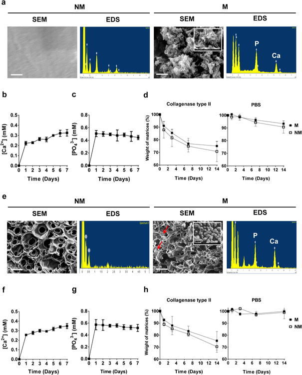
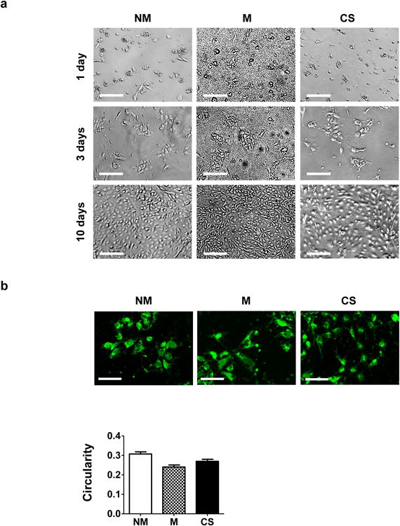
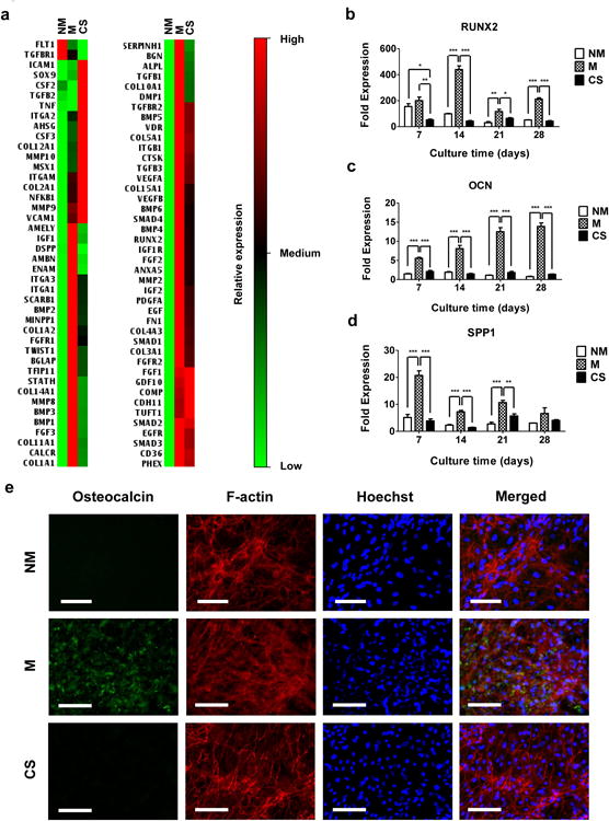
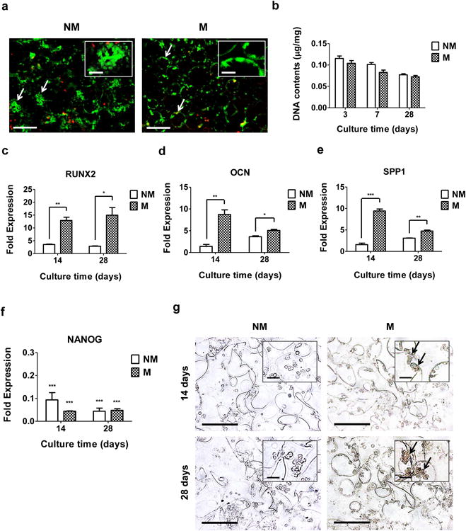
Human induced pluripotent stem cells (hiPSC) are a promising cell source with pluripotency and self-renewal properties. Design of simple and robust biomaterials with an innate ability to induce lineage-specificity of hiPSC is desirable to realize their application in regenerative medicine. In this study, the potential of biomaterials containing calcium phosphate minerals to induce osteogenic differentiation of hiPSC was investigated. hiPSC cultured using mineralized gelatin methacrylate-based matrices underwent osteogenic differentiation ex vivo, in both two-dimensional and three-dimensional cultures, in growth medium devoid of any osteogenic-inducing chemical components or growth factors. The findings that osteogenic differentiation of hiPSC can be achieved through biomaterial-based cues alone present new avenues for personalized regenerative medicine. Such biomaterials that could not only act as structural scaffolds, but could also provide tissue-specific functions such as directing stem cell differentiation commitment, have great potential in bone tissue engineering.

Keywords: Bone tissue engineering; Calcium phosphate; Gelatin methacrylate; Human induced pluripotent stem cells; Osteogenic differentiation.


Conflict of interest statement

Conflict of interest: Authors declare no conflict of interest.

Figures

Figure 1
Figure 1. Development and characterization of GelMA-based matrices
(a) SEM images and corresponding EDS for non-mineralized (NM) and mineralized (M) gelatin-methacrylate-co-acryloyl 6-aminocaprioic acid (GelMA-co-A6ACA) hydrogels. Scale bar: 1 μm. Inset shows high magnification image. Scale bar: 200 nm. Dissolution of (b) Ca2+ and (c) PO43- from mineralized GelMA-co-A6ACA hydrogels incubated in Tris-HCl buffer at 37 °C as a function of time. (d) In vitro degradation of non-mineralized and mineralized GelMA-co-A6ACA hydrogels in 0.02 w/v% collagenase type II solution or PBS at 37 °C as a function of time. (e) SEM images and corresponding EDS for non-mineralized and mineralized gelatin-methacrylate-co-acryloyl 6-aminocaproic acid-co-poly(ethylene glycol)-diacrylate (GelMA-co-A6ACA-co-PEGDA) macroporous hydrogels. Red arrows indicate mineral structures. Scale bar: 100 μm. Inset shows high magnification image. Scale bar: 1 μm. Release of (f) Ca2+ and (g) PO43- from mineralized GelMA-co-A6ACA-co-PEGDA macroporous hydrogels in Tris-HCl buffer at 37 °C as a function of time. (h) In vitro degradation of non-mineralized and mineralized GelMA-co-A6ACA-co-PEGDA macroporous hydrogels in 0.02 w/v% collagenase type II solution or PBS at 37 °C as a function of time. Data are presented as mean ± standard errors (n=3).
Figure 2
Figure 2. Attachment and proliferation of hiPSCs on different matrices
(a) Bright field images for hiPSCs after 1, 3, and 10 days of culture on non-mineralized (NM) and mineralized (M) hydrogels and gelatin-coated coverslips (CS). Scale bar: 200 μm. (b) Images of hiPSCs labeled using CellTracker after 3 days of culture on non-mineralized and mineralized hydrogels and coverslips. Scale bar: 100 μm. The bar graph shows the quantitative representation of the circularity; the circularity indices were determined from the stained images. Data are shown as mean ± standard errors (n=30).
Figure 3
Figure 3. Osteogenic differentiation of hiPSCs on CaP-rich mineralized hydrogels in 2-D culture
(a) Gene expression array analyses of hiPSCs cultured for 28 days on non-mineralized (NM) and mineralized (M) hydrogels and coverslips (CS). Relative expressions: red (high), black (medium), and green (low). Gene expressions of (b) RUNX2, (c) OCN, and (d) SPP1 for hiPSCs cultured on non-mineralized and mineralized hydrogels and coverslips as a function of culture time. Data are presented as fold expression of target genes after normalization to undifferentiated, pluripotent hiPSCs. (e) Immunofluorescent staining for OCN (green) and F-actin (red) of hiPSCs cultured on non-mineralized and mineralized hydrogels and coverslips for 28 days. Nuclei are stained blue with Hoechst. Scale bars represent 100 μm. Data are displayed as mean ± standard errors (n=3). (b-d) Comparison of multiple groups at the same time point was made by one-way ANOVA with Tukey-Kramer post-hoc test. Asterisks indicate statistical significances corresponding to p-values (*: p < 0.05; **: p < 0.01; ***: p < 0.001).
Figure 4
Figure 4. Osteogenic differentiation of hiPSCs on CaP-rich mineralized macroporous hydrogels in 3-D culture
(a) Live-dead staining of hiPSC-laden non-mineralized (NM) and mineralized (M) macroporous hydrogels after 3 days of culture. Arrows and inset indicate aggregated and spread hiPSCs within the non-mineralized and mineralized matrices, respectively. Scale bars represent 200 μm and scale bars in the inset indicate 50 μm. (b) DNA contents of hiPSCs cultured using non-mineralized and mineralized matrices as a function of culture time. Data are presented as DNA contents after normalization to dry weight of matrices. Gene expression of (c) RUNX2, (d) OCN, (e) SPP1, and (f) NANOG of hiPSCs on non-mineralized and mineralized matrices as a function of culture time. Data are presented as fold expression of target genes after normalization to undifferentiated, pluripotent hiPSCs. (g) Immunohistochemical staining for OCN of hiPSCs on non-mineralized and mineralized matrices as a function of culture time. Scale bars represent 100 μm. Inset shows high magnification images. Arrows indicate positive stains. Scale bars in the inset represent 20 μm. Data are displayed as mean ± standard errors (n=3). (c-e) Two groups at the same time point were compared by two-tailed Student's t-test. (f) All the groups were compared to undifferentiated, pluripotent hiPSCs prior to their culture on all matrices by two-way ANOVA with Bonferroni post-hoc test. Asterisks represent statistical significances according to p-values. (*: p < 0.05; **: p < 0.01; ***: p < 0.001).

References

    1. Amabile G, Meissner A. Induced pluripotent stem cells: current progress and potential for regenerative medicine. Trends in molecular medicine. 2009;15:59–68. - PubMed
    1. Wobus AM, Boheler KR. Embryonic stem cells: prospects for developmental biology and cell therapy. Physiological reviews. 2005;85:635–78. - PubMed
    1. Takahashi K, Tanabe K, Ohnuki M, Narita M, Ichisaka T, Tomoda K, et al. Induction of pluripotent stem cells from adult human fibroblasts by defined factors. cell. 2007;131:861–72. - PubMed
    1. Teng S, Liu C, Krettek C, Jagodzinski M. The Application of Induced Pluripotent Stem Cells for Bone Regeneration: Current Progress and Prospects. Tissue Engineering Part B: Reviews. 2013 - PubMed
    1. Illich DJ, Demir N, Stojković M, Scheer M, Rothamel D, Neugebauer J, et al. Concise review: induced pluripotent stem cells and lineage reprogramming: prospects for bone regeneration. Stem Cells. 2011;29:555–63. - PubMed

Publication types

MeSH terms

LinkOut - more resources