Review
. 2015 Jun;30(6):903-10.
doi: 10.1093/ndt/gfu281. Epub 2014 Aug 25.

Who regenerates the kidney tubule?

Affiliations
Review

Who regenerates the kidney tubule?

Rafael Kramann et al. Nephrol Dial Transplant. 2015 Jun.

Abstract

The kidney possesses profound regenerative potential and in some cases can recover completely 'restitutio at integrum' following an acute kidney injury (AKI). Emerging evidence strongly suggests that sometimes repair is incomplete, however, and, in this situation, an episode of AKI leads to future chronic kidney disease (CKD). Understanding the tubular response after AKI will shed light on the relationship between incomplete repair and future risk of CKD. The first repair phase after AKI is characterized by robust proliferation of epithelial cells in the proximal tubule. The exact source of these proliferating cells has been a source of controversy for the last decade. While nearly everyone now agrees that reparative cells arise within the proximal tubule, there is disagreement about whether all surviving cells possess an equivalent repair capacity through dedifferentiation, or alternatively whether a pre-existing intratubular stem cell population [so-called scattered tubular cells (STC)] is responsible for repair. This review will summarize the evidence on both sides of this issue and will discuss very recent genetic fate-tracing data that strongly points against the existence of intratubular stem cells but rather indicates that terminally differentiated proximal tubule epithelial cells undergo dedifferentiation upon injury to replace lost neighboring tubular epithelial cells through proliferative self-duplication. This new evidence includes data clearly indicating that STC are not committed tubular stem cells but instead represent individual dedifferentiated tubular epithelial cells that transiently express putative stem cell markers.

Keywords: acute kidney injury; dedifferentiation; stem cell; tubular repair.


Figures

FIGURE 1:
FIGURE 1:
Rationale of clonal and dilution analysis using SLC34a1GFPCreER (SLC34a1GCE/+), Rosa26tomato/+ mice. (A) We first induce single-cell labeling in terminally differentiated proximal tubular epithelial cells by very low-dose tamoxifen injection into SLC34a1GCE/+ mice. Therefore, on a clone size–frequency curve, nearly all clones exist as single-cell clones (blue curve in both panels). If a stem cell population contributes to renal repair, the clone size will remain single and clone frequency will decrease after injury, due to death of differentiated cells after injury (green curve in left panel). If repair is by self-duplication of terminally differentiated cells (= labeled cells), the clone size will expand through proliferation of single-cell clones during repair (red curve in right panel), resulting in a rightward shift of the clone size–frequency curve. (B) To determine whether an intratubular stem cell contributes to repair at all, we performed dilution analysis. Complete cell labeling in the terminally differentiated proximal tubular was accomplished by high-dose tamoxifen administration. A round of IRI and repair follows. Upon analysis of repaired tubules, if only terminally differentiated cells contribute to renal repair, the labeling will remain undiluted by unlabeled cells (Option 1). If an unlabeled progenitor population contributes to repair of the genetic label will be diluted by progeny of these cells (Option 2). Our results support Option 1, self-duplication, as the only mechanism of proximal tubule repair.
FIGURE 2:
FIGURE 2:
Clonal and dilution analysis of SLC34a1GCE/+, Rosa26tomato/+ mice. (A) In uninjured contralateral kidney, cells were labeled solely by low-dose tamoxifen (left panel). In the IRI kidney, clone size of labeled cells expanded after repair (right panel). (B) Dilution analysis of SLC34a1GCE/+, Rosa26tomato/+ mice. These representative images indicate complete labeling of the proximal tubule in uninjured kidneys and remaining labeling (no dilution) after injury. Staining for BrdU (green) indicates the proliferating cells of the proximal tubule indeed came from the labeled (red) terminally differentiated tubular cells. (C and D) Immunostaining of dedifferentiation markers; vimentin and Pax2. In non-injured kidneys terminally differentiated labeled epithelial cells (red) did never stain for vimentin or Pax2. Whereas, following injury (IRI) these terminally differentiated cells became flattened and expressed both vimentin and Pax2, indicating dedifferentiation.
FIGURE 3:
FIGURE 3:
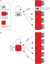
Dueling models for epithelial repair after injury. (A) In the stem/progenitor model, scattered progenitors are located adjacent to differentiated proximal tubule epithelium. The progenitors express CD24, CD133, Kim-1 and Vimentin, among other markers. After injury, these cells preferentially survive and selectively proliferate. Their progeny differentiate into proximal tubular epithelial cells. (B) In the self-duplication model, any fully differentiated cell that survives the injury has an equivalent capacity to dedifferentiate and proliferate. The progeny then re-differentiate into proximal tubular tubule epithelium. In this case, CD24, CD133, Kim-1 and vimentin are not markers of a separate stem cell compartment, but are injury markers transiently expressed by a dedifferentiated epithelial cell. Thus, new epithelia derive from their fully differentiated neighbors in a process of self-duplication.

References

    1. Basile DP, Anderson MD, Sutton TA. Pathophysiology of acute kidney injury. Compr Physiol 2012; 2: 1303–1353 - PMC - PubMed
    1. Coca SG, Singanamala S, Parikh CR. Chronic kidney disease after acute kidney injury: a systematic review and meta-analysis. Kidney Int 2012; 81: 442–448 - PMC - PubMed
    1. Uchida S, Endou H. Substrate specificity to maintain cellular ATP along the mouse nephron. Am J Physiol 1988; 255(5 Pt 2): F977–F983 - PubMed
    1. Lieberthal W, Nigam SK. Acute renal failure. I. Relative Importance of Proximal vs. Distal Tubular Injury. Am J Physiol 1998; 275(5 Pt 2): F623–F631 - PubMed
    1. Bonventre JV, Yang L. Cellular pathophysiology of ischemic acute kidney injury. J Clin Invest 2011; 121: 4210–4221 - PMC - PubMed

Publication types

MeSH terms