. 2014 Sep 8;24(17):2025-32.
doi: 10.1016/j.cub.2014.07.038. Epub 2014 Aug 21.

Actin is required for IFT regulation in Chlamydomonas reinhardtii

Affiliations

Actin is required for IFT regulation in Chlamydomonas reinhardtii

Prachee Avasthi et al. Curr Biol. .

Abstract

Assembly of cilia and flagella requires intraflagellar transport (IFT), a highly regulated kinesin-based transport system that moves cargo from the basal body to the tip of flagella [1]. The recruitment of IFT components to basal bodies is a function of flagellar length, with increased recruitment in rapidly growing short flagella [2]. The molecular pathways regulating IFT are largely a mystery. Because actin network disruption leads to changes in ciliary length and number, actin has been proposed to have a role in ciliary assembly. However, the mechanisms involved are unknown. In Chlamydomonas reinhardtii, conventional actin is found in both the cell body and the inner dynein arm complexes within flagella [3, 4]. Previous work showed that treating Chlamydomonas cells with the actin-depolymerizing compound cytochalasin D resulted in reversible flagellar shortening [5], but how actin is related to flagellar length or assembly remains unknown. Here we utilize small-molecule inhibitors and genetic mutants to analyze the role of actin dynamics in flagellar assembly in Chlamydomonas reinhardtii. We demonstrate that actin plays a role in IFT recruitment to basal bodies during flagellar elongation and that when actin is perturbed, the normal dependence of IFT recruitment on flagellar length is lost. We also find that actin is required for sufficient entry of IFT material into flagella during assembly. These same effects are recapitulated with a myosin inhibitor, suggesting that actin may act via myosin in a pathway by which flagellar assembly is regulated by flagellar length.


Figures

Figure 1
Figure 1
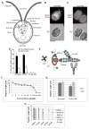
The actin polymerization inhibitor Latrunculin B causes flagellar shortening. (A) Diagram of a Chlamydomonas cell with relevant or prominent organelles identified. (B) Representative images showing Lifeact-Venus localization to the mid-cell portion as filaments and to the cell-anterior region as patches (arrowheads) and (C) its complete loss upon treatment with latrunculin B. Bar 5μm. (D) Lifeact-Venus expressing cells were cultured to OD = 0.3 and treated with 0.1% DMSO or 10 μM latrunculin B. 100–250 cells were counted by fluorescence microscopy for localization of Lifeact-Venus at the mid-cell portion and as cell-anterior patches. The error bars indicate 95% confidence intervals from three replicates. * and ** indicate p < 0.05 and 0.01, respectively. (E) Diagram of point of action for inhibitors. Latrunculin B (LatB) binds actin monomers to prevent filament assembly (1); SMIFH2 inhibits Formin mediated actin nucleation (2); CK-666 inhibits Arp2/3 mediated branched actin assembly (3); Cytochalasin D (CD) binds barbed ends of actin filaments (4) with high affinity and G-actin (1) with low affinity; jasplakinolide (jasplak) binds F-actin at the interface of 3 actin subunits (5); blebbistatin binds myosin motor (6). (F) Treatment with latrunculin B to impair actin polymerization and ultimately disassemble the actin network results in a dose dependent decrease in flagellar length. Concentrations indicated correspond to the markers on the line graph. (G) Treatment with 10μM latrunculin B shortens wild-type flagella but not ida5 mutant flagella demonstrating that the flagellar shortening effect is caused by targeting actin and not a nonspecific effect of impaired cell health (*:p< .00000005). (H) Inhibiting an actin nucleation factor, formin, with SMIFH2 or inhibiting actin branch promoting Arp2/3 with CK-666, shortens flagellar length in a dose dependent manner. KVSM18 and CK-689 are inactive controls for SMIFH2 and CK-666 respectively and have no effect. 1% DMSO are also included as controls as inhibitors are diluted in DMSO. DMSO 1 refers to the control for the formin experiment and DMSO 2 refers to the control for the Arp2/3 experiment. Error bars are 95% confidence intervals. Asterisks indicate significant difference from controls (p<0.05). See also Figure S1.
Figure 2
Figure 2
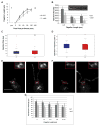
Flagellar length, IFT train size and basal body accumulation of IFT material are perturbed during flagellar regeneration in a mutant lacking conventional actin. (A) Flagellar regeneration following pH shock mediated flagellar shedding results in a rapid initial phase of growth and slow late phase as flagella near final length in wild type cells. In ida5 actin mutants, initial phase of growth is much slower but ultimately flagellar length equals wild type. Error bars are 95% confidence intervals. (*:p<0.0005, **:p<0.00005)(B) As flagella regenerate, wild-type cells show typical IFT injection behavior with larger trains injected in shorter flagella and smaller trains injected in longer flagella. ida5 mutants retain length-dependent injection of IFT material but the injection sizes are significantly smaller than wild-type (p<0.05 for each bin). Means and standard error are shown. Inset is an example of the type of kymograph data that is analyzed for this graph. Vertical scale bar is 5μm. Horizontal scale bar is 5 sec. (C) Box and whisker plot of injection sizes shows median (black bar), 25th and 75th percentiles (box top and bottom edges), extreme data points not considered outliers (whiskers) and outliers (red crosses). There is no significant difference (p=0.6581) in injection sizes between wild type and ida5 mutants at steady state. (D) Box and whisker plot of injection frequency shows no significant difference between wild type and ida5 mutants (p=0.1214). Injection frequency is the inverse of the measured wait time. (E) Projection of z-stacks taken of a representative image for KAP-GFP expressing cells. 0.2μm step TIFFs were used for image analysis for Figure 3C. Red box shows region surrounding basal bodies. Scale bar is 5μm. Insets are 3x magnifications. Left panel shows short flagella early in regeneration. Right panel shows full length flagella late in regeneration. (F) Projection of z-stacks of a representative image for KAP-GFP expressing cells on ida5 mutant background. Left panel shows short flagella early in regeneration. Right panel shows full length flagella late in regeneration. (G) Intensity of KAP-GFP motor fluorescence at basal bodies is reduced in ida5 mutants compared to wild type initially, mirroring the initial decrease in flagellar regeneration kinetics. Wild type accumulation of KAP-GFP is known to decrease with flagellar length, as less IFT material is required when flagellar growth slows. When growth slows and ida5 flagella reach wild-type lengths (7–12 microns), ida5 mutant accumulation of KAP-GFP at basal bodies is no longer statistically significantly different from wild-type levels. Asterisks indicate significant difference from wild type (p<0.05). See also Figure S2.
Figure 3
Figure 3
Myosin inhibition alters flagellar assembly kinetics. (A) In contrast to the deceleratory kinetics seen in wild-type flagellar regeneration, cells regenerating in the presence of the active (−)-blebbistatin enantiomer regeneration at a roughly constant rate. All error bars are 95% confidence intervals. (*:p<0.05, **:p<0.005, ***:p<0.0005, ****:p<0.00005, *****:p<0.000005) (B) For regeneration in inactive (+)-blebbistatin enantiomer, kymograph analysis of fluorescence intensity of IFT trains injected into flagella shows typical wild type behavior. As flagella elongate, IFT trains injected become smaller. In contrast, IFT injection sizes are reduced (*:p<.005) and do not significantly decrease with flagellar length in active (−)-blebbistatin. (C) Unlike inactive control, accumulation of KAP-GFP at basal bodies is also reduced (*:p<0.005) at shorter lengths and flagellar length-independent in (−)-blebbistatin, suggesting that myosin-II activity is required for length-dependent mobilization of IFT material for flagellar assembly. Error bars are the standard error of the mean. (D) Superimposition of Dictyostelium myosin II (PDB ID: 1YV3) crystallized with bound (−)-blebbistatin (purple), chicken myosin Va (PDB ID: 1W7J, cyan), and the homology model of putative Chlamydomonas myosin VIII (Phytozyme ID Myo2/Cre09.g416250.t1), with (−)-blebbistatin docked (orange). The loop containing Leu262 in myosin II moves away to allow (−)-blebbistatin to bind; in myosin V, the homologous Leu243 completely obstructs (−)-blebbistatin binding. All three putative Chlamydomonas myosins also have a leucine in this position. In myosin II, Ser456 is small enough to not encroach the (−)-blebbistatin binding site. However, the homologous bulky Tyr439 substitution forces not only the entire loop to move away from the binding site, but it also forces ATP, here represented by the BeF3 ion, to bind lower than the VO43− ion in myosin II. All three putative Chlamydomonas myosins also have a tyrosine in this position. See also Figure S3.
Figure 4
Figure 4
Myo2-Venus shows similar localization to LifeAct-Venus; this localization is sensitive to latrunculin B and is transiently lost during regeneration. (A) Representative images showing Myo2-Venus localization to the mid-cell portion as filaments and to the cell-anterior region as dots (arrowheads). (B) Only the mid-cell localization was lost upon latrunculin B addition. Bar, 5 μm. (C) Myo2-Venus expressing cells were cultured and counted as in (Figure 1D). * and **** indicate p < 0.05 and 0.0001, respectively. (D) Loss of mid-cell Myo2 localization during initial phase of flagellar regeneration. 100 cells were counted at each time point. Error bars indicate 95% confidence intervals from three replicates. *, **, and *** indicates p < 0.05, 0.01, and 0.001, respectively. Bar, 5 μm. See also Figure S4.

Comment in

References

    1. Pedersen LB, Rosenbaum JL. Intraflagellar transport (IFT) role in ciliary assembly, resorption and signalling. Curr Top Dev Biol. 2008;85:23–61. - PubMed
    1. Ludington WB, Wemmer KA, Lechtreck KF, Witman GB, Marshall WF. Avalanche-like behavior in ciliary import. Proc Natl Acad Sci U S A. 2013;110:3925–3930. - PMC - PubMed
    1. Hirono M, Uryu S, Ohara A, Kato-Minoura T, Kamiya R. Expression of conventional and unconventional actins in Chlamydomonas reinhardtii upon deflagellation and sexual adhesion. Eukaryotic Cell. 2003;2:486–493. - PMC - PubMed
    1. Kato-Minoura T, Uryu S, Hirono M, Kamiya R. Highly divergent actin expressed in a Chlamydomonas mutant lacking the conventional actin gene. Biochem Biophys Res Commun. 1998;251:71–76. - PubMed
    1. Dentler WL, Adams C. Flagellar microtubule dynamics in Chlamydomonas: cytochalasin D induces periods of microtubule shortening and elongation; and colchicine induces disassembly of the distal, but not proximal, half of the flagellum. J Cell Biol. 1992;117:1289–1298. - PMC - PubMed

Publication types

MeSH terms

Substances