. 2014 Oct;94(2):367-82.
doi: 10.1111/mmi.12770. Epub 2014 Sep 15.

Structure of the Mycobacterium tuberculosis type VII secretion system chaperone EspG5 in complex with PE25-PPE41 dimer

Affiliations

Structure of the Mycobacterium tuberculosis type VII secretion system chaperone EspG5 in complex with PE25-PPE41 dimer

Natalia Korotkova et al. Mol Microbiol. 2014 Oct.

Abstract

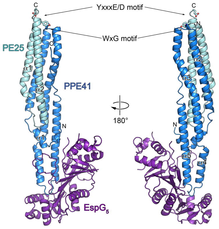
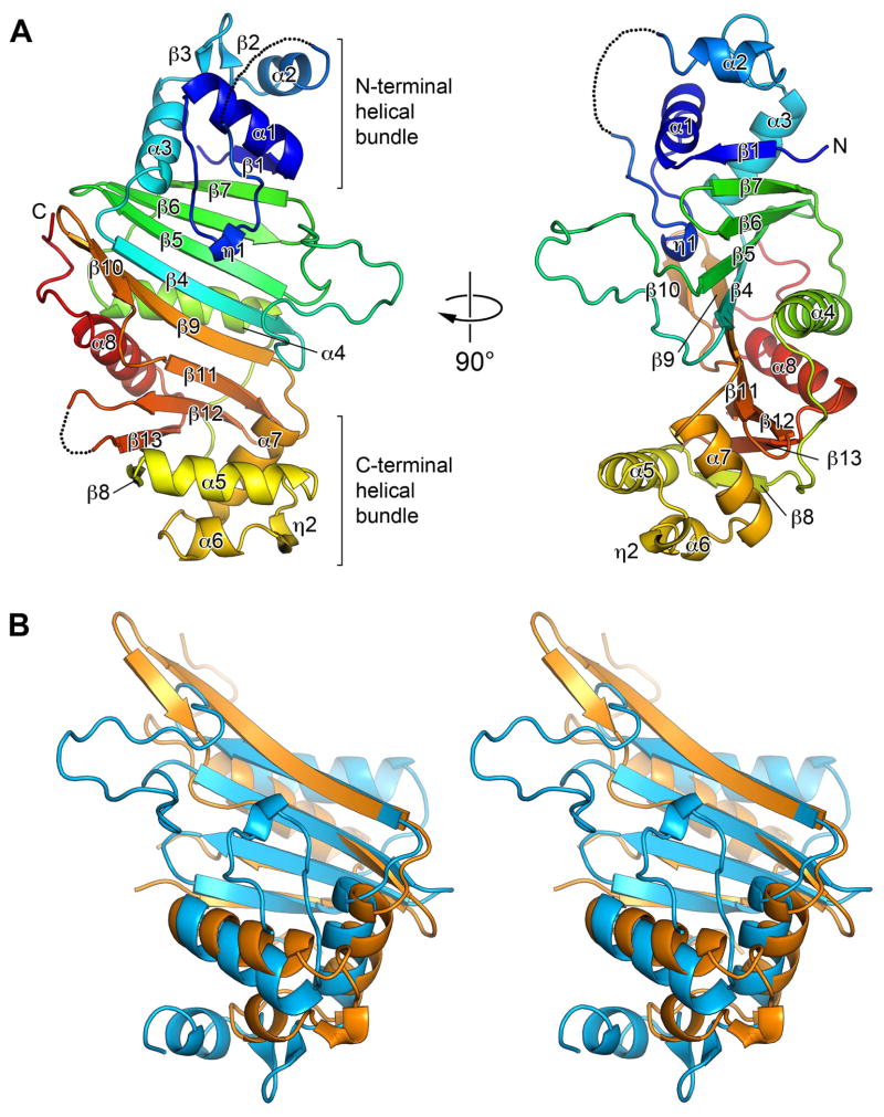
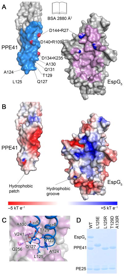
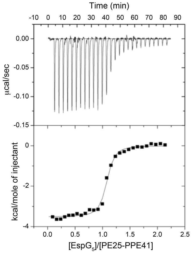
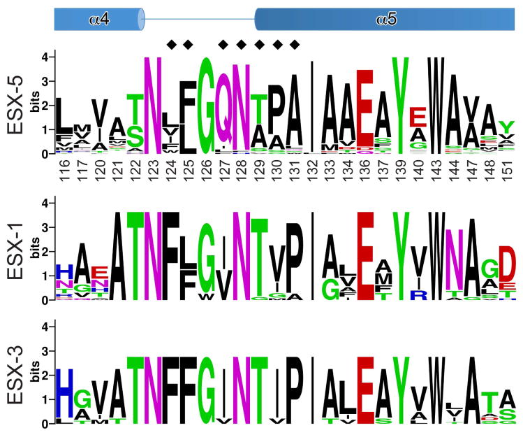
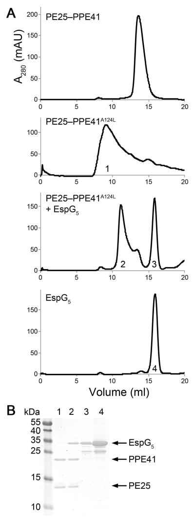
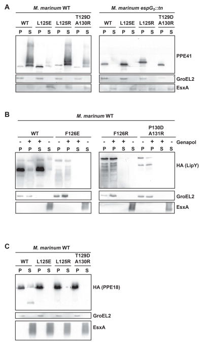
The growth or virulence of Mycobacterium tuberculosis bacilli depends on homologous type VII secretion systems, ESX-1, ESX-3 and ESX-5, which export a number of protein effectors across membranes to the bacterial surface and environment. PE and PPE proteins represent two large families of highly polymorphic proteins that are secreted by these ESX systems. Recently, it was shown that these proteins require system-specific cytoplasmic chaperones for secretion. Here, we report the crystal structure of M. tuberculosis ESX-5-secreted PE25-PPE41 heterodimer in complex with the cytoplasmic chaperone EspG(5). EspG(5) represents a novel fold that is unrelated to previously characterized secretion chaperones. Functional analysis of the EspG(5) -binding region uncovered a hydrophobic patch on PPE41 that promotes dimer aggregation, and the chaperone effectively abolishes this process. We show that PPE41 contains a characteristic chaperone-binding sequence, the hh motif, which is highly conserved among ESX-1-, ESX-3- and ESX-5-specific PPE proteins. Disrupting the interaction between EspG(5) and three different PPE target proteins by introducing different point mutations generally affected protein secretion. We further demonstrate that the EspG(5) chaperone plays an important role in the ESX secretion mechanism by keeping aggregation-prone PE-PPE proteins in their soluble state.


Conflict of interest statement

Conflict of interest

The authors declare that they have no conflict of interest.

Figures

Fig. 1
Fig. 1. Crystal structure of the M. tuberculosis PE25–PPE41–EspG5 complex
Ribbon representation of the complex in two views related by ~180° rotation. EspG5 interacts with PPE protein on the opposite side from the conserved YxxxD/E and WxG motifs of PE25–PPE41 dimer.
Fig. 2
Fig. 2. Structure of EspG5 represents a novel fold with quasi 2-fold symmetry
A. Ribbon representation is colored in rainbow colors from N-terminus (blue) to C-terminus (red). Secondary structure elements are labeled α1–α8 and β1–β13. The disordered loops are indicated as dashed lines. The two views are related by ~90° rotation. B. Stereo view of the structural superposition of the N-terminal (blue) and the C-terminal (orange) sub-domains of EspG5.
Fig. 3
Fig. 3. The interface between EspG5 and PPE41
A. An ‘open book’ view of the PPE41–EspG5 complex. Contact residues in the interface are colored in light blue (PPE41) and purple (EspG5). Atoms participating in intermolecular salt bridges are colored in red and blue. B. EspG5 and PPE41 are shown in the same orientation as in panel (A). The surface is colored according to electrostatic surface potential contoured at ±5 kT e−1, with red corresponding to a negative and blue to a positive potential. The contact areas are indicated by black lines. C. EspG5 is shown in surface representation as in panel (B), PPE41 is shown in ribbon representation (blue). Residues in α4-α5 loop are shown in stick representation. Hydrogen bonds are shown as black dashed lines. D. SDS-PAGE analysis of co-purification of EspG5 and PE25–PPE41 mutant variant dimers. Proteins were purified by affinity chromatography from the lysate of E. coli strain expressing N-terminally His-tagged PE25, PPE41 and EspG5. Note that PPE41L125E has an altered mobility on SDS-PAGE.
Fig. 4
Fig. 4. Isothermal calorimetry of PE25–PPE41 with EspG5
EspG5 (100 μM) was injected into a solution of PE25–PPE41 (10 μM) containing the same buffer. The resulting isotherm data were fit to a single-site model, which gave an average dissociation constant of 48.1 nM. One representative data set is shown here.
Fig. 5
Fig. 5. EspG5-binding region of PPE41
The sequence conservation of the EspG5-binding site is displayed as a sequence logo (http://weblogo.berkeley.edu) (Crooks et al., 2004) based on the sequence alignments of ESX-5- and ESX-3-specific PPE proteins of M. tuberculosis H37Rv and ESX-1-specific PPE68 homologs from mycobacteria (Fig. S2, S6 and S7). Secondary structure elements of PPE41 are shown at the top. Black diamonds indicate residues that were subjected to mutational analysis (Table 2 and Fig. 3C, S8, S9 and S10).
Fig. 6
Fig. 6. EspG5 binding to PE25–PPE41A124L mutant complex induces dis-aggregation of the heterodimer
A. Size-exclusion chromatography of PE25–PPE41, PE25–PPE41A124L mutant, EspG5 incubated with PE25–PPE41A124L mutant and EspG5. B. SDS-PAGE analysis of the peak fractions indicated (1–4) from the size-exclusion chromatography.
Fig. 7
Fig. 7. Effect of the substitutions in the PPE41 interface on PE25–PPE41 secretion in M. marinum
A. Immunoblot analysis of PPE41 in culture supernatant and cell pellet fractions of M. marinum WT (left panel) and its EspG5 transposon mutant (right panel). Equivalent amounts of protein were loaded. GroEL and EsxA were used as a cytoplasmic and secreted fraction controls, respectively. Analysis of LipY (panel B) and PPE18 (panel C) in culture supernatant and cell pellet fractions of M. marinum WT.

References

    1. Abdallah AM, Verboom T, Hannes F, Safi M, Strong M, Eisenberg D, et al. A specific secretion system mediates PPE41 transport in pathogenic mycobacteria. Mol Microbiol. 2006;62:667–679. - PubMed
    1. Abdallah AM, Verboom T, Weerdenburg EM, Gey van Pittius NC, Mahasha PW, Jimenez C, et al. PPE and PE_PGRS proteins of Mycobacterium marinum are transported via the type VII secretion system ESX-5. Mol Microbiol. 2009;73:329–340. - PubMed
    1. Arbing MA, Kaufmann M, Phan T, Chan S, Cascio D, Eisenberg D. The crystal structure of the Mycobacterium tuberculosis Rv3019c–Rv3020c ESX complex reveals a domain-swapped heterotetramer. Protein Sci. 2010;19:1692–1703. - PMC - PubMed
    1. Barathy DV, Suguna K. Crystal structure of a putative aspartic proteinase domain of the Mycobacterium tuberculosis cell surface antigen PE_PGRS16. FEBS Open Bio. 2013;3:256–262. - PMC - PubMed
    1. Birtalan SC, Phillips RM, Ghosh P. Three-dimensional secretion signals in chaperone-effector complexes of bacterial pathogens. Mol Cell. 2002;9:971–980. - PubMed

Publication types

MeSH terms

LinkOut - more resources