. 2014 Sep;144(3):221-30.
doi: 10.1085/jgp.201411223.

K⁺ channel gating: C-type inactivation is enhanced by calcium or lanthanum outside

Affiliations

K⁺ channel gating: C-type inactivation is enhanced by calcium or lanthanum outside

Clay M Armstrong et al. J Gen Physiol. 2014 Sep.

Abstract

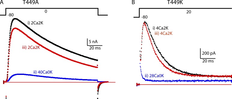
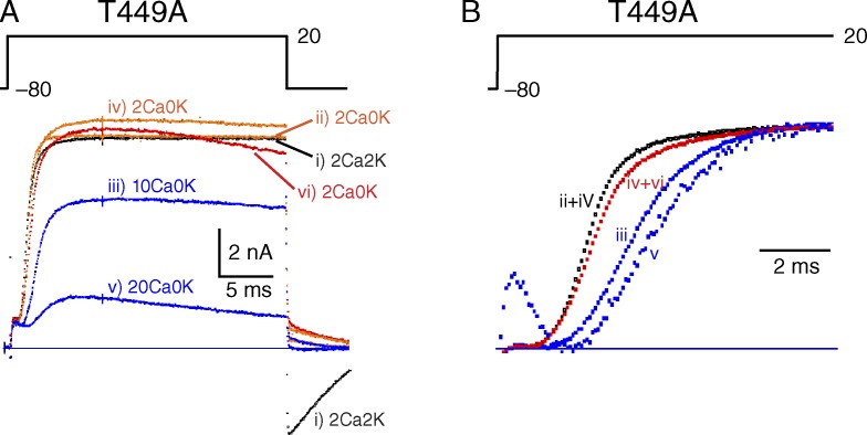
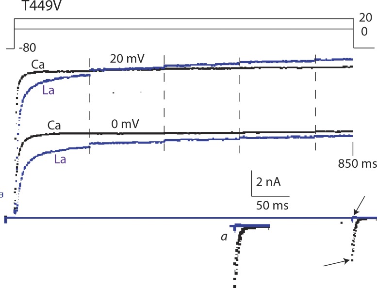
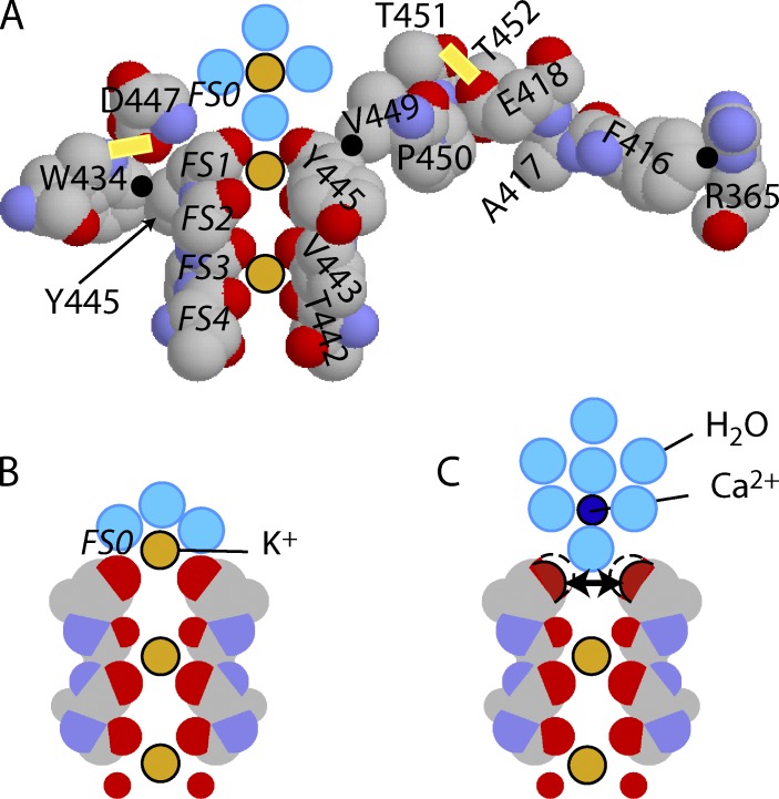
Many voltage-gated K(+) channels exhibit C-type inactivation. This typically slow process has been hypothesized to result from dilation of the outer-most ring of the carbonyls in the selectivity filter, destroying this ring's ability to bind K(+) with high affinity. We report here strong enhancement of C-type inactivation upon extracellular addition of 10-40 mM Ca(2+) or 5-50 µM La(3+). These multivalent cations mildly increase the rate of C-type inactivation during depolarization and markedly promote inactivation and/or suppress recovery when membrane voltage (V(m)) is at resting levels (-80 to -100 mV). At -80 mV with 40 mM Ca(2+) and 0 mM K(+) externally, ShBΔN channels with the mutation T449A inactivate almost completely within 2 min or less with no pulsing. This behavior is observed only in those mutants that show C-type inactivation on depolarization and is distinct from the effects of Ca(2+) and La(3+) on activation (opening and closing of the V(m)-controlled gate), i.e., slower activation of K(+) channels and a positive shift of the mid-voltage of activation. The Ca(2+)/La(3+) effects on C-type inactivation are antagonized by extracellular K(+) in the low millimolar range. This, together with the known ability of Ca(2+) and La(3+) to block inward current through K(+) channels at negative voltage, strongly suggests that Ca(2+)/La(3+) acts at the outer mouth of the selectivity filter. We propose that at -80 mV, Ca(2+) or La(3+) ions compete effectively with K(+) at the channel's outer mouth and prevent K(+) from stabilizing the filter's outer carbonyl ring.


Figures

Figure 1.
Figure 1.
In two mutants that have C-type inactivation, high extracellular Ca2+ depresses IK. (A) The traces show IK from a cell expressing T449A during a depolarization lasting 110 ms from −80 to 0 mV. The traces were taken in the order i–iii, with a rest of ∼2 min between them to allow full recovery from C-type inactivation. IK is strongly depressed by 40Ca0K. Note, however, that the time constant of inactivation of the current remaining in 40Ca0K is similar to that in 2Ca2K. (B) IK traces from a T449K cell. The extracellular solution was changed from 4Ca2K (i) to 28Ca0K (ii) and then back to 4Ca2K (iii).
Figure 2.
Figure 2.
Ca2+ does not appreciably inhibit currents through T449V channels. (A) Whole-cell currents elicited by pulses from −80 to 50 mV in 4Ca2K and 40Ca0K. (B) Slower activation time course of T449V in 40Ca0K (blue) compared with those in 4Ca2K (black). (C) The currents for pulses from −80 to 0 mV are scaled by 1.3 times to account for the familiar right shift of the I-V curve caused by high Ca2+, which is shown in D. (Gilly and Armstrong, 1982; Hille, 2001). All results shown are from the same cell.
Figure 3.
Figure 3.
Ca2+ action on C-type inactivation examined with short pulses. (A) A cell expressing T449A was stimulated at 0.1 Hz in several solutions, beginning with 2Ca2K. In each solution, maximum IK was in a steady-state after ∼10 pulses or less, and a current record for the 11th pulse in each solution is displayed. The currents were elicited by 25-ms pulses from −80 to 20 mV and then back to −80 mV. (B) Slowing of activation kinetics by 10 and 20 Ca2+. The currents from A are scaled vertically for comparison and displayed on a fast time scale. ii+iv, iv+vi: mean of ii and iv or iv and vi, respectively. A gating current transient is visible in trace v, the most amplified.
Figure 4.
Figure 4.
Enhancement of C-type inactivation by high extracellular Ca2+ is antagonized by extracellular K+. (A) Whole-cell currents from a T449A cell were elicited by 25-ms voltage pulses from −80 to 20 mV applied at 0.1 Hz. Peak outward current in nanoamperes is plotted as a function of time as Ca2+ and K+ concentrations in the extracellular solutions were changed as indicated at the top. Solution changes were completed over the course of several pulses. The numbers (1–7) are explained in the section C-type inactivation examined with short pulses. (B) Whole-cell currents from a cell expressing T449A channels during exposure to 40Ca0K or 40Ca2K. The currents were elicited by pulses from −80 to 50 mV after a rest of 2 min.
Figure 5.
Figure 5.
Extracellular La3+ enhances C-type inactivation in T449A and T449T (i.e., wild type) channels. (A) The black and the red trace are IK from an outside-out patch before and after exposure to 50 µM La3+, which virtually eliminated IK (blue trace). IK was elicited by 1.5-s pulses from –90 to 0 mV every 62 s. (B) Peak IK through T449A channels before and during exposure to 5 µM La3+ in the extracellular solution. Similar results were obtained in four patches. (C) Peak IK through T449T (wild type) channels before, during, and after washout of 5 µM La3+ in the extracellular solution. The patch was held at −90 mV except to measure the currents as illustrated in the insets. Similar results were obtained in four patches. The extracellular solution contained (mM) 130 NaCl, 4 KCl, 2 CaCl2, 2 MgCl2, 10 glucose, and 10 HEPES, pH 7.4, plus 5 µM La3+ in blue traces. The internal solution contained (mM) 140 KCl, 2 MgCl2, and 10 HEPES, pH 7.2.
Figure 6.
Figure 6.
Extracellular La3+ (10 µM) slows activation of T449V channels but causes no inactivation. IK is compared in 10KCa2K (black traces) and in 10Ca2K + La3+ (10 µM La3+; blue traces). Sampling was interrupted at each of the vertical dashed lines for 100 ms and then resumed. Total sweep duration was 850 ms. On return to −80 mV, there was a clear inward IK tail without La3+(bottom arrow), but none in the presence of La3+, which blocks K+ channels at negative voltage (top arrow). The tail is shown at slightly higher gain in inset a.
Figure 7.
Figure 7.
K+ occupancy of FS1 is affected by voltage and extracellular Ca2+/La3+. (A) A slice through the selectivity filter and some surrounding residues. Points of significant close contact between residues are shown by black dots. Two significant H-bonds are indicated by the yellow bars. The spheres on the pore axis are dehydrated K+ ions in filter sites FS1 and FS3 and a hydrated K+ in FS0, just outside the filter (water molecules are light blue). (B) The filter only, in occupancy state FS2–FS4. When conducting, occupancy alternates between FS1–FS3 and FS2–FS4. At rest, when the channel is closed and Vm is negative, FS2–FS4 is the preferred state. In this state, the outermost carbonyls are stabilized by attraction to a partially dehydrated K+ ion in FS0. (C) FS0 is occupied by a Ca2+ ion. Its hydration shell is too firmly attached to allow close approach to the outer carbonyls of FS1. As a result, they repel, as indicated by the two-headed arrow, leading to dilation and C-type inactivation. Residue numbering is appropriate to ShBΔN.

References

    1. Åqvist, J., and Luzhkov V.. 2000. Ion permeation mechanism of the potassium channel. Nature. 404:881–884 10.1038/35009114 - DOI - PubMed
    1. Armstrong, C.M.1971. Interaction of tetraethylammonium ion derivatives with the potassium channels of giant axons. J. Gen. Physiol. 58:413–437 10.1085/jgp.58.4.413 - DOI - PMC - PubMed
    1. Baukrowitz, T., and Yellen G.. 1995. Modulation of K+ current by frequency and external [K+]: a tale of two inactivation mechanisms. Neuron. 15:951–960 10.1016/0896-6273(95)90185-X - DOI - PubMed
    1. Burgess, J.1978. Metal Ions in Solution. Ellis Horwood, Chichester, NY: 481 pp
    1. Campbell, D.T., and Hille B.. 1976. Kinetic and pharmacological properties of the sodium channel of frog skeletal muscle. J. Gen. Physiol. 67:309–323 10.1085/jgp.67.3.309 - DOI - PMC - PubMed

Publication types