. 2014 Sep 17;136(37):12899-911.
doi: 10.1021/ja507614f. Epub 2014 Sep 8.

A phosphohistidine proteomics strategy based on elucidation of a unique gas-phase phosphopeptide fragmentation mechanism

Affiliations

A phosphohistidine proteomics strategy based on elucidation of a unique gas-phase phosphopeptide fragmentation mechanism

Rob C Oslund et al. J Am Chem Soc. .

Abstract

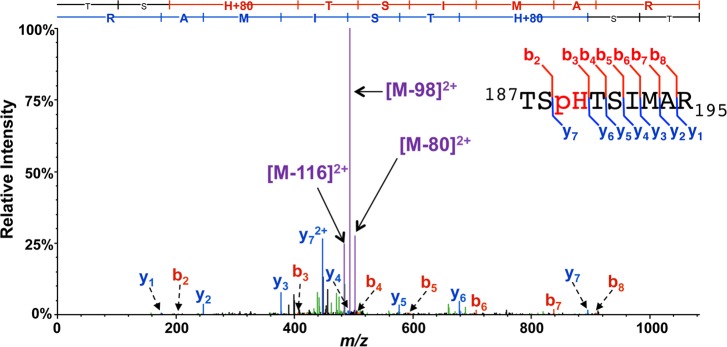
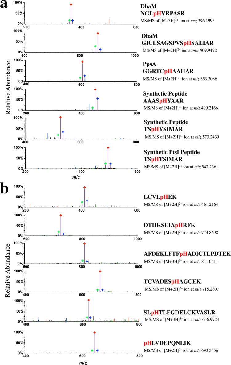
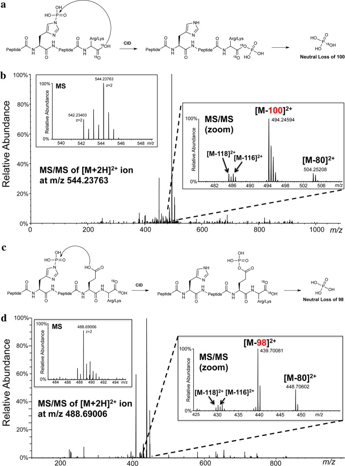
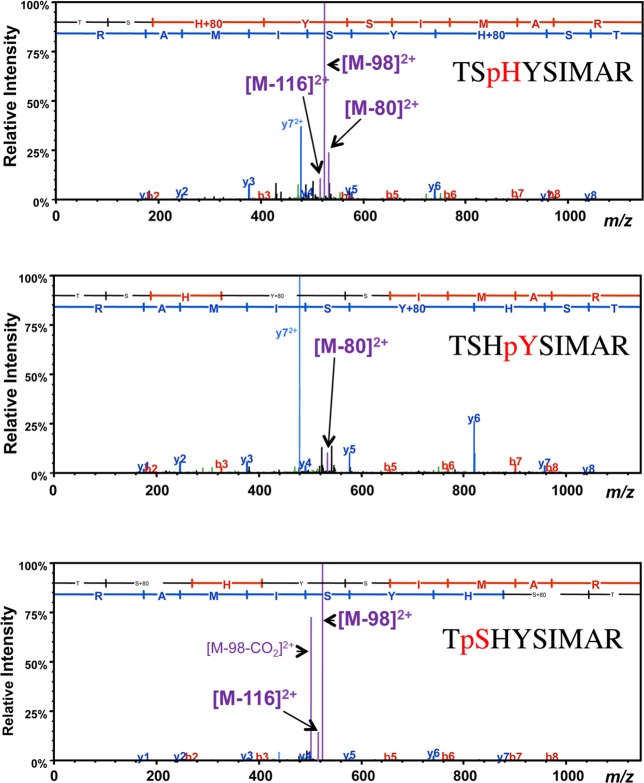
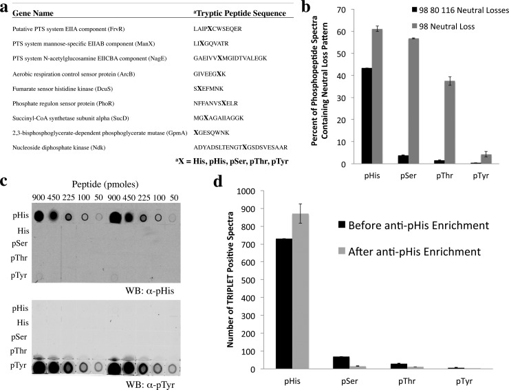
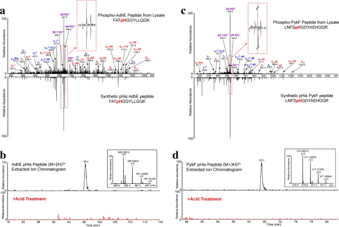
Protein histidine phosphorylation is increasingly recognized as a critical posttranslational modification (PTM) in central metabolism and cell signaling. Still, the detection of phosphohistidine (pHis) in the proteome has remained difficult due to the scarcity of tools to enrich and identify this labile PTM. To address this, we report the first global proteomic analysis of pHis proteins, combining selective immunoenrichment of pHis peptides and a bioinformatic strategy based on mechanistic insight into pHis peptide gas-phase fragmentation during LC-MS/MS. We show that collision-induced dissociation (CID) of pHis peptides produces prominent characteristic neutral losses of 98, 80, and 116 Da. Using isotopic labeling studies, we also demonstrate that the 98 Da neutral loss occurs via gas-phase phosphoryl transfer from pHis to the peptide C-terminal α-carboxylate or to Glu/Asp side chain residues if present. To exploit this property, we developed a software tool that screens LC-MS/MS spectra for potential matches to pHis-containing peptides based on their neutral loss pattern. This tool was integrated into a proteomics workflow for the identification of endogenous pHis-containing proteins in cellular lysates. As an illustration of this strategy, we analyzed pHis peptides from glycerol-fed and mannitol-fed Escherichia coli cells. We identified known and a number of previously speculative pHis sites inferred by homology, predominantly in the phosphoenolpyruvate:sugar transferase system (PTS). Furthermore, we identified two new sites of histidine phosphorylation on aldehyde-alcohol dehydrogenase (AdhE) and pyruvate kinase (PykF) enzymes, previously not known to bear this modification. This study lays the groundwork for future pHis proteomics studies in bacteria and other organisms.


Figures

Figure 1
Figure 1
MS/MS spectrum of a pHis tryptic peptide, TSpHTSIMAR, from the endogenously phosphorylated E. coli protein PtsI. MS/MS was performed by linear ion trap CID, during which prominent species derived from the neutral loss of 98, 80, and 116 Da dominate the ion current. Major b- and y-ion backbone fragments and species derived from neutral losses off the precursor (M) are indicated in red, blue, and purple, respectively. Inset into the spectrum is a summary sequence, flagged to indicate the detected b- and y-ions, also shown in the sequence ladder above the spectrum.
Figure 2
Figure 2
Neutral losses of 98, 80, and 116 Da in CID MS/MS spectra of diverse pHis peptides. Prominent neutral loss triplets (Δ98, Δ80, and Δ116 Da) were observed in the CID MS/MS spectra for (a) tryptic peptides from known pHis proteins (DhaM, PpsA) and chemically phosphorylated synthetic peptides, respectively, and (b) pHis peptides derived from chemically phosphorylated BSA. The Δ116, Δ98, and Δ80 Da product ion species are indicated in purple with each peak labeled with a green, red, or blue diamond, respectively. Precursor peptide sequences, charge state, and m/z values are shown to the right of each spectrum. MS/MS spectra showing the full recorded m/z range for these CID experiments are shown in Figure S1.
Figure 3
Figure 3
Neutral loss of 98 Da from pHis peptides occurs predominantly through the C-terminal or a side chain carboxylate. (a) Proposed model showing loss of phosphoric acid from pHis peptides via the C-terminal carboxylate during CID. (b) CID MS/MS of the [M + 2H]2+ pHis peptide TSpHTSIMAR-C18O18OH ion showing the prominent neutral loss of 100 Da. Inset into the spectrum is a high resolution MS spectrum of the precursor species at m/z 544.23763 (left panel) and a high resolution MS/MS spectrum showing species corresponding to the primary loss of 100 Da and less prominent losses of 80, 118, and 116 Da (right panel). (c) Proposed model showing loss of phosphoric acid from pHis peptides via a side chain carboxylate during CID. (d) CID MS/MS of the [M + 2H]2+ pHis peptide SpHEFMNK-C18O18OH ion showing the prominent neutral loss of 98 Da. Inset into the spectrum is a high resolution MS spectrum of the precursor species at m/z 488.69006 (left panel) and a high resolution MS/MS spectrum showing species corresponding to the primary loss of 98 Da and less prominent losses of 80, 118, and 116 Da (right panel).
Figure 4
Figure 4
CID Fragmentation of pHis, pTyr, and pSer peptides display distinct neutral loss patterns. CID MS/MS spectra of a family of phosphopeptides containing the same underlying peptide sequence, TSHYSIMAR. The pHis peptide TSpHYSIMAR exhibited the distinct CID-induced neutral loss pattern of Δ98, Δ80 and Δ116 Da. This triplet neutral loss pattern was not observed for the isobaric pSer (TpSHYSIMAR) and pTyr (TSHpYSIMAR) peptides. b- and y-ions and neutral loss species are indicated in the spectra, as in Figure 1. Precursor peptide sequences are as labeled.
Figure 5
Figure 5
Specificity of a second-generation pHis antibody and its use for pHis peptide immunoprecipitation in combination with triplet neutral loss filtering for highly selective discrimination of pHis from pSer/pThr/pTyr peptide analogues. (a) Phosphopeptide library sequences, consisting of tryptic peptides containing the known pHis sites of 9 E. coli proteins. The library was diversified by replacing the pHis site with pSer, pThr, pTyr, or His residues. (b) Library peptides were analyzed by CID MS/MS, and the percentage of individual MS/MS spectra displaying the Δ98, Δ80, and Δ116 Da neutral loss pattern (black bars) or the Δ98 neutral loss (gray bars) was calculated for each phosphotype. Note that these neutral losses were not observed for any of the nonphosphorylated peptides (not shown). (c) Dot blot analysis of the library of phosphopeptides in (a), pooled by phosphotype, using a newly developed α-pHis antibody and an α-pTyr antibody (4G10, Millipore) (see Figure S14 for loading control). (d) Library peptides from (a) were analyzed by CID MS/MS before and after immunoprecipitation with the α-pHis antibody. Tallies of MS/MS spectra displaying the triplet neutral loss pattern were generated for each of the library constituents by the TRIPLET software, and the results were grouped according to pHis, pSer, pThr, or pTyr phosphotype.
Figure 6
Figure 6
Validation of AdhE and PykF pHis MS/MS peptide assignments. (a) CID MS/MS spectrum of the endogenous E. coli AdhE pHis peptide annotated with the major matched b- and y-type ions indicated. The synthetic Adhe pHis peptide is shown in the mirror image. Inset into the spectrum is an enlargement of the neutral loss pattern for the endogenous vs the synthetic pHis peptide. (b) Extracted ion chromatogram of the [M + 2H]2+ AdhE pHis peptide ion from E. coli lysate (top) and from the acid-treated lysate (bottom, red trace). Inset into the spectrum is a high resolution MS spectrum of the precursor species at m/z 686.32679. (c) CID MS/MS spectrum of the endogenous E. coli PykF pHis peptide, with the prominent matched b- and y-type ions as indicated. The synthetic PykF pHis peptide is shown in the mirror image. Inset into the spectrum is an enlargement of the neutral loss pattern for the endogenous vs the synthetic pHis peptide. (d) Extracted ion chromatogram of the [M + 3H]3+ PykF pHis peptide ion from E. coli lysate (top) and from the acid-treated lysate (bottom, red trace). Inset into the spectrum is a high resolution MS spectrum of the precursor species at m/z 570.90574.

References

    1. Walsh C. T.Posttranslational Modification of Proteins: Expanding Nature’s Inventory; Roberts and Company Publishers: Greenwood Village, CO, 2006.
    1. Cohen P. Nat. Rev. Drug Discovery 2002, 1, 309. - PubMed
    1. Dephoure N.; Gould K. L.; Gygi S. P.; Kellogg D. R. Mol. Biol. Cell 2013, 24, 535. - PMC - PubMed
    1. Kee J.-M.; Muir T. W. ACS Chem. Biol. 2012, 7, 44. - PMC - PubMed
    1. Attwood P. V.; Besant P. G.; Piggott M. J. Amino Acids 2010, 40, 1035. - PubMed

Publication types

MeSH terms