. 2014 Nov;82(11):4718-28.
doi: 10.1128/IAI.02198-14. Epub 2014 Aug 25.

Synergistic interactions of Pseudomonas aeruginosa and Staphylococcus aureus in an in vitro wound model

Affiliations

Synergistic interactions of Pseudomonas aeruginosa and Staphylococcus aureus in an in vitro wound model

Stephanie DeLeon et al. Infect Immun. 2014 Nov.

Abstract

In individuals with polymicrobial infections, microbes often display synergistic interactions that can enhance their colonization, virulence, or persistence. One of the most prevalent types of polymicrobial infection occurs in chronic wounds, where Pseudomonas aeruginosa and Staphylococcus aureus are the two most common causes. Although they are the most commonly associated microbial species in wound infections, very little is known about their interspecies relationship. Evidence suggests that P. aeruginosa-S. aureus coinfections are more virulent than monoculture infection with either species; however, difficulties in growing these two pathogens together in vitro have hampered attempts to uncover the mechanisms involved. Here we describe a simple and clinically relevant in vitro wound model that supported concomitant growth of P. aeruginosa and S. aureus. We observed that the ability of P. aeruginosa and S. aureus to survive antibiotic treatment increased when they were grown together in planktonic cocultures and that antibiotic tolerance was further enhanced when they were grown together in the wound model. We attributed this enhanced tolerance to both the "host-derived" and "bacterium-derived" matrix components. Taken together, our data indicate that P. aeruginosa and S. aureus may benefit each other by coinfecting wounds and that the host-derived matrix may serve as important a role as the bacterium-derived matrix in protecting bacteria from some antibiotics.


Figures

FIG 1
FIG 1
P. aeruginosa and S. aureus can be grown together in a “wound-like” environment. (A to C) Coagulated WLM supports the growth of P. aeruginosa and S. aureus (A), which are clearly discernible and in close proximity within the magnified area (B, inset) of a thin section of coagulated WLM stained with H&E (B) or DAPI (C). (D) Clusters of P. aeruginosa and S. aureus can be visualized in H&E-stained thin sections from mouse wounds. Visualization was carried out with a UPlan FL 40× oil objective (numerical aperture, 1.30).
FIG 2
FIG 2
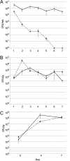
S. aureus was quickly eliminated in planktonic LB cocultures but not in static wound environments. (A and B) S. aureus–P. aeruginosa cocultures were initiated in LB medium (A) or WLM (B) with approximately 104 CFU of each species and were grown in glass culture tubes under static, aerobic conditions at 37°C for 7 days. Cultures were sampled at the indicated time points, and the numbers of bacteria were estimated by CFU enumeration on P. aeruginosa and S. aureus isolation media. Experiments were performed in triplicate. Dashed lines, S. aureus; solid lines, P. aeruginosa. (C) S. aureus–P. aeruginosa coinfections were initiated in mouse surgical excision wounds (6 mice per time point) with approximately 104 CFU of each species (P. aeruginosa and S. aureus), and equal numbers were maintained over 7 days.
FIG 3
FIG 3
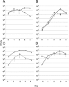
P. aeruginosa and S. aureus reached equilibrium in the in vitro wound environment. Regardless of whether cocultures were initiated in WLM with a high inoculum (105 CFU/species) (A, C, and D) or a low inoculum (102 CFU/species) (B), or with a P. aeruginosa/S. aureus starting ratio of 1:1 (B), 100:1 (C), or 1:100 (D), they reached similar maximum bacterial loads after growth in glass culture tubes under static, aerobic conditions at 37°C for 4 days. Cultures were sampled at the indicated time points, and the numbers of bacteria were estimated by CFU enumeration on P. aeruginosa and S. aureus isolation media. Experiments were performed in triplicate. Dashed lines, S. aureus; solid lines, P. aeruginosa.
FIG 4
FIG 4
Coculturing P. aeruginosa and S. aureus altered their antibiotic susceptibilities. Planktonic monocultures or cocultures of P. aeruginosa (PA) and S. aureus (SA) were grown overnight in WLM, and tolerances to gentamicin, ciprofloxacin, and tetracycline were determined. Cultures were initiated with a ∼1:1 starting ratio of P. aeruginosa to S. aureus and were grown in flasks at 37°C with vigorous shaking, which prevented the coagulation of the medium. One-way ANOVA and the Tukey-Kramer multiple-comparison test were used to test for differences in tolerance between S. aureus and P. aeruginosa monocultures and cocultures. Each group included at least 6 individual cultures. Error bars represent the standard errors of the means.
FIG 5
FIG 5
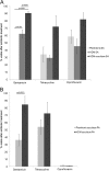
The host-derived matrix contributed to the gentamicin tolerance of S. aureus (A) and P. aeruginosa (B). S. aureus monocultures and S. aureus–P. aeruginosa cocultures were grown aerobically overnight at 37°C either in culture tubes under static conditions, which allows for the coagulation of the WLM, or in flasks with vigorous shaking, which inhibits coagulation and results in a planktonic population. The gentamicin, tetracycline, and ciprofloxacin tolerances of samples from these cultures were then measured. One-way ANOVA and the Tukey-Kramer multiple-comparison test were used to test for differences in tolerance to each antibiotic group between S. aureus cultures. Two-tailed, unpaired t tests were used to test for differences in tolerance to each antibiotic group between planktonic and HDM-associated P. aeruginosa cocultures. Each group included at least 6 individual cultures. Error bars represent the standard errors of the means.
FIG 6
FIG 6
Imaging of P. aeruginosa and S. aureus in the HDM. (A and B) One-day-old P. aeruginosa–S. aureus cocultures in coagulated WLM were frozen in a cryomatrix and were sectioned. Sections were stained with FITC-conjugated ConA and DAPI and were visualized by fluorescence microscopy (with UPlan FL 40× [A] and 100× [B] oil objectives [numerical aperture, 1.30]). These images revealed discrete clusters of bacteria, which stained intensely for ConA, interspersed in the fibrous host matrix. (C) Scanning electron microscopy (magnification, ×15,000), performed on glutaraldehyde- and paraformaldehyde-fixed sections from 1-day-old P. aeruginosa–S. aureus cocultures in coagulated WLM, revealed cocci and rods in close proximity enmeshed in a web of fibrous material.
FIG 7
FIG 7
P. aeruginosa and S. aureus biofilm mutants demonstrated reduced levels of tolerance in the wound environment. The gentamicin tolerance levels of wild-type (w/t) S. aureus and P. aeruginosa were compared to those of isogenic mutants with mutations in EPS-related genes. Strains were grown in coagulated WLM (A) or in murine surgical excision wounds (B). (A) One-way ANOVA and the Tukey-Kramer multiple-comparison test were used to test for differences in tolerance among strains grown in vitro. Each group included at least 6 individual cultures. Error bars represent the standard errors of the means. (B) A paired t test was used to test for significant differences in tolerance between P. aeruginosa or S. aureus strains grown in vivo (6 mice per group; error bars represent the standard errors of the means).

References

    1. Wolcott RD, Rhoads DD, Bennett ME, Wolcott BM, Gogokhia L, Costerton JW, Dowd SE. 2010. Chronic wounds and the medical biofilm paradigm. J. Wound Care 19:45–46, 48–50, 52–53. 10.12968/jowc.2010.19.2.46966. - DOI - PubMed
    1. Cunningham AB. 2006. Biofilms: the hypertextbook. Montana State University, Bozeman, MT.
    1. James GA, Swogger E, Wolcott R, Pulcini E, Secor P, Sestrich J, Costerton JW, Stewart PS. 2008. Biofilms in chronic wounds. Wound Repair Regen. 16:37–44. 10.1111/j.1524-475X.2007.00321.x. - DOI - PubMed
    1. Dowd SE, Sun Y, Secor PR, Rhoads DD, Wolcott BM, James GA, Wolcott RD. 2008. Survey of bacterial diversity in chronic wounds using pyrosequencing, DGGE, and full ribosome shotgun sequencing. BMC Microbiol. 8:43. 10.1186/1471-2180-8-43. - DOI - PMC - PubMed
    1. Singh VA, Barbul A. 2008. Bacterial biofilms in wounds. Wound Repair Regen. 16:1. 10.1111/j.1524-475X.2007.00349.x. - DOI - PubMed

Publication types

MeSH terms

Substances