. 2014 Nov;82(11):4729-45.
doi: 10.1128/IAI.01876-14. Epub 2014 Aug 25.

Localization of Burkholderia cepacia complex bacteria in cystic fibrosis lungs and interactions with Pseudomonas aeruginosa in hypoxic mucus

Affiliations

Localization of Burkholderia cepacia complex bacteria in cystic fibrosis lungs and interactions with Pseudomonas aeruginosa in hypoxic mucus

Ute Schwab et al. Infect Immun. 2014 Nov.

Abstract

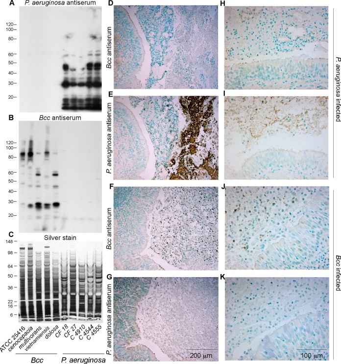
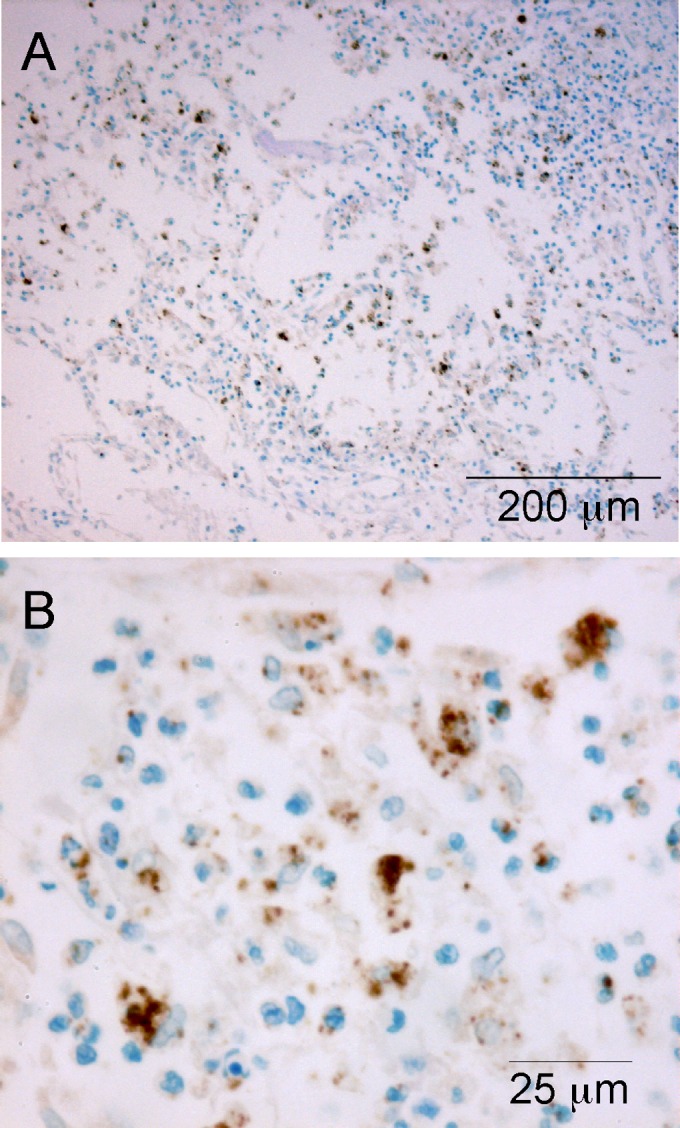
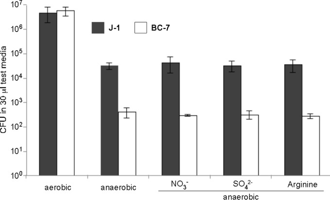
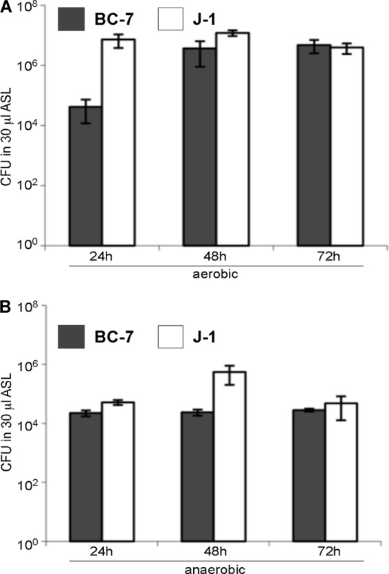
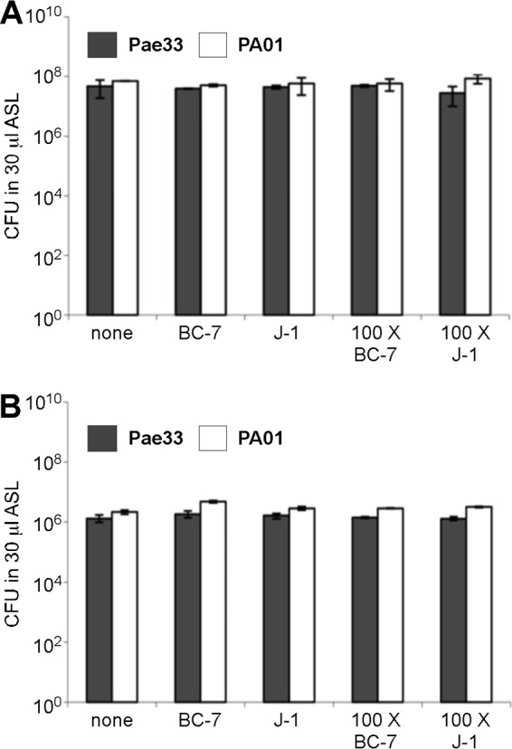
The localization of Burkholderia cepacia complex (Bcc) bacteria in cystic fibrosis (CF) lungs, alone or during coinfection with Pseudomonas aeruginosa, is poorly understood. We performed immunohistochemistry for Bcc and P. aeruginosa bacteria on 21 coinfected or singly infected CF lungs obtained at transplantation or autopsy. Parallel in vitro experiments examined the growth of two Bcc species, Burkholderia cenocepacia and Burkholderia multivorans, in environments similar to those occupied by P. aeruginosa in the CF lung. Bcc bacteria were predominantly identified in the CF lung as single cells or small clusters within phagocytes and mucus but not as "biofilm-like structures." In contrast, P. aeruginosa was identified in biofilm-like masses, but densities appeared to be reduced during coinfection with Bcc bacteria. Based on chemical analyses of CF and non-CF respiratory secretions, a test medium was defined to study Bcc growth and interactions with P. aeruginosa in an environment mimicking the CF lung. When test medium was supplemented with alternative electron acceptors under anaerobic conditions, B. cenocepacia and B. multivorans used fermentation rather than anaerobic respiration to gain energy, consistent with the identification of fermentation products by high-performance liquid chromatography (HPLC). Both Bcc species also expressed mucinases that produced carbon sources from mucins for growth. In the presence of P. aeruginosa in vitro, both Bcc species grew anaerobically but not aerobically. We propose that Bcc bacteria (i) invade a P. aeruginosa-infected CF lung when the airway lumen is anaerobic, (ii) inhibit P. aeruginosa biofilm-like growth, and (iii) expand the host bacterial niche from mucus to also include macrophages.


Figures

FIG 1
FIG 1
Specificity of P. aeruginosa and Bcc antisera and control immunohistochemistry. (A to C) Replicate blots of bacterial lysates (loaded as indicated along the bottom) stained with P. aeruginosa and Bcc antisera (A and B), as indicated, and a replicate silver-stained gel (C) to assess loading. (D to K). Bronchial sections from CF individuals whose sputum was positive for B. cenocepacia and negative for P. aeruginosa (patient 1) (Table 1), or vice versa (patient 21), were stained with Bcc and P. aeruginosa antisera. Low-magnification (D to G) and higher-magnification (H to K) images illustrate that the antisera were specific for their respective bacteria. Variable staining of the ciliated epithelial cell luminal border and diffuse faint staining of connective tissue matrix were observed with both antisera and were considered nonspecific. Double the concentration of normal rabbit serum typically displayed negligible staining (not shown).
FIG 2
FIG 2
Localization of B. cenocepacia and P. aeruginosa in CF lungs removed during transplantation. (A) Low-power view of a bronchus from a 32-year-old male whose last preoperative sputum culture was positive for B. cenocepacia and negative for P. aeruginosa (patient 10). The luminal mucopurulent secretions react strongly with Bcc antiserum, with more densely stained areas but without obvious colonial associations. (B) Higher-power view of panel A illustrating predominantly intracellular material in phagocytes and occasional intercellular individual bacteria or pairs/small clusters of bacteria. (C) Low-power view of a bronchus from a 28-year-old female (patient 21) (Table 1) whose last preoperative sputum culture was positive for P. aeruginosa and negative for Bcc bacteria. There are abundant ball-like bacterial colonies and sheet-like masses in the luminal secretions that react strongly with P. aeruginosa antiserum. (D) Higher-power view from panel C illustrating bacterial colonies.
FIG 3
FIG 3
Localization of B. cenocepacia in a CF lung obtained at autopsy. (A) Section of distal lung parenchyma from a 20-year-old male (patient 1) (Table 1) whose prior sputum culture was positive for B. cenocepacia and negative for P. aeruginosa. Highly inflamed alveoli contain cells and particulate matter that react strongly with Bcc antiserum. (B) Higher-power view from panel A, again illustrating predominantly intracellular material in phagocytes and occasional intercellular individual bacteria or pairs/small clusters of bacteria.
FIG 4
FIG 4
Aerobic and anaerobic growth of Bcc bacteria in modified M9 medium. To test for strategies used by Bcc bacteria to gain energy anaerobically, we developed a test medium (modified M9 medium), which was supplemented with the electron acceptors nitrate, sulfate, and arginine. B. multivorans J-1 and B. cenocepacia BC-7 (approximately 200 to 500 CFU/1 μl PBS) were incubated for 72 h under aerobic and anaerobic conditions in 30 μl of test medium (n = 3), and the number of bacteria was determined by serial dilutions and plating.
FIG 5
FIG 5
Aerobic and anaerobic growth of Bcc bacteria in in vitro ASL harvested from well-differentiated airway cultures. B. multivorans J-1 and B. cenocepacia BC-7 (approximately 200 to 500 CFU/1 μl PBS) were incubated for 24, 48, and 72 h under aerobic (A) and anaerobic (B) conditions in 30 μl of in vitro ASL (n = 3), and the number of bacteria was determined by serial dilutions and plating.
FIG 6
FIG 6
Growth of Bcc bacteria in mucin gels. Purified mucins from bovine cervical mucus were suspended in PBS, and the mucin gels were incubated with B. multivorans J-1 and B. cenocepacia BC-7 (approximately 200 to 500 CFU/1 μl PBS) for 72 h under anaerobic conditions (n = 3). Enumeration was performed by serial dilutions (see also Fig. S1 in the supplemental material).
FIG 7
FIG 7
Degradation of mucins by bacterial cell lysates. Purified bovine cervical mucins were incubated with trypsin (Trp) or crude lysates from B. cenocepacia (BC-7) or B. multivorans (J-1), and the products were resolved by 1% agarose gel electrophoresis (A and B) or 4-to-20% gradient PAGE (C to E). (A) The agarose gel was vacuum blotted and probed with a mucin-specific antibody. (B) The intensity profile for each lane of panel A was determined by using the linescan function in MetaMorph. Lines were extended vertically down each lane, and the average pixel intensity in each row of pixels across the width of the lane was recorded. (C) The PAGE gel was stained with PAS stain to reveal glycosylated materials, which correspond to mucins. Band 1 represents intact mucins (Con) in the stacking gel or fragments thereof that are too large to penetrate the running gel. Bands 2 and 3 represent glycosylated materials in the running gel that resulted from digestion. (D) Control PAGE gel showing the resolution of glycosylated materials in the bacterial lysate in the absence of exogenously added mucins. Note that bacterial cell lysates contained no band corresponding to mucin degradation products. (E) Intensity profiles of the lanes in panel C, as described above for panel B.
FIG 8
FIG 8
Competitive growth of Bcc and P. aeruginosa bacteria in in vitro ASL at 48 h under aerobic conditions. Equal numbers (A) or a 100:1 ratio (B) of B. multivorans J-1 or B. cenocepacia BC-7 and P. aeruginosa Pae33 or PAO1 bacteria was incubated in in vitro ASL under aerobic conditions for 48 h (n = 3). Samples were removed, serial dilutions were performed, and samples in parallel were plated onto Trypticase soy agar and Burkholderia cepacia selective agar (BCSA) plates for enumeration.
FIG 9
FIG 9
Competitive growth of Bcc and P. aeruginosa bacteria in in vitro ASL at 72 h under anaerobic conditions. Equal numbers (A) or a 100:1 ratio (B) of B. multivorans J-1 or B. cenocepacia BC-7 and P. aeruginosa Pae33 or PAO1 bacteria was incubated in in vitro ASL under anaerobic conditions for 72 h (n = 3). Samples were removed at specific time points, serial dilutions were performed, and samples in parallel were plated onto Trypticase soy agar and BCSA plates for enumeration.
FIG 10
FIG 10
Aerobic and anaerobic growth of P. aeruginosa in the presence and absence of Bcc bacteria in in vitro ASL. Equal numbers or a 100:1 ratio of B. multivorans J-1 or B. cenocepacia BC-7 and P. aeruginosa Pae33 or PAO1 bacteria was incubated in ASL under aerobic (A) and anaerobic (B) conditions for 72 h (n = 3). Samples were removed at specific time points, serial dilutions were performed, and samples in parallel were plated onto Trypticase soy agar and BCSA plates for enumeration.

Similar articles

Cited by

References

    1. Cystic Fibrosis Foundation. 2013. Cystic Fibrosis Foundation patient registry: 2012 annual data report. Cystic Fibrosis Foundation, Bethesda, MD.
    1. Mahenthiralingam E, Urban TA, Goldberg JB. 2005. The multifarious, multireplicon Burkholderia cepacia complex. Nat. Rev. Microbiol. 3:144–156. 10.1038/nrmicro1085. - DOI - PubMed
    1. Frangolias DD, Mahenthiralingam E, Rae S, Raboud JM, Davidson AGF, Wittmann R, Wilcox PG. 1999. Burkholderia cepacia in cystic fibrosis—variable disease course. Am. J. Respir. Crit. Care Med. 160:1572–1577. - PubMed
    1. Nielsen AT, Tolker-Nielsen T, Barken KB, Molin S. 2000. Role of commensal relationships on the spatial structure of a surface-attached microbial consortium. Environ. Microbiol. 2:59–68. 10.1046/j.1462-2920.2000.00084.x. - DOI - PubMed
    1. Riedel K, Hentzer M, Geisenberger O, Huber B, Steidle A, Wu H, Høiby N, Givskov M, Molin S, Eberl L. 2001. N-Acylhomoserine-lactone-mediated communication between Pseudomonas aeruginosa and Burkholderia cepacia in mixed biofilms. Microbiology 147:3249–3262. - PubMed

Publication types

MeSH terms