. 2014 Nov;82(11):4789-801.
doi: 10.1128/IAI.02279-14. Epub 2014 Aug 25.

Differential regulation of proinflammatory cytokine expression by mitogen-activated protein kinases in macrophages in response to intestinal parasite infection

Affiliations

Differential regulation of proinflammatory cytokine expression by mitogen-activated protein kinases in macrophages in response to intestinal parasite infection

Mei Xing Lim et al. Infect Immun. 2014 Nov.

Abstract

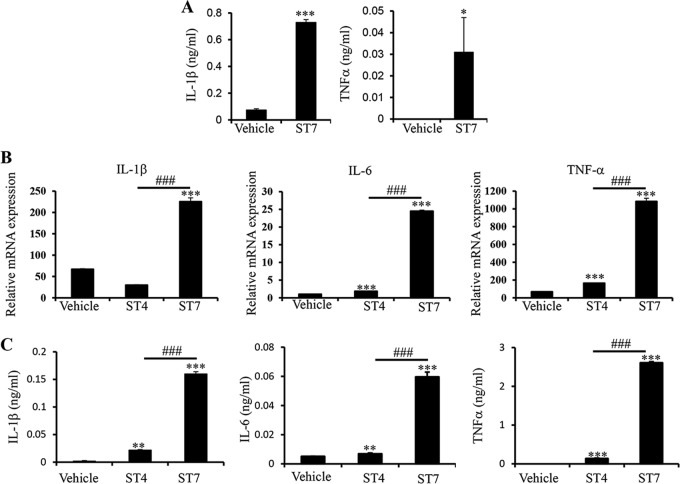
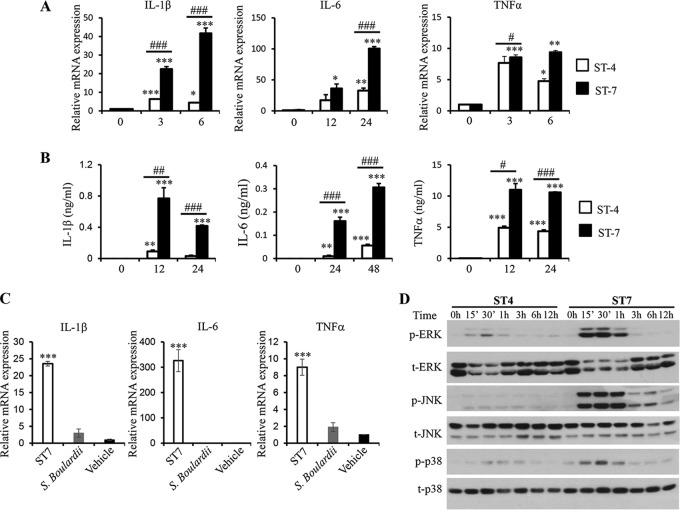
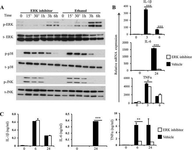
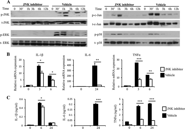
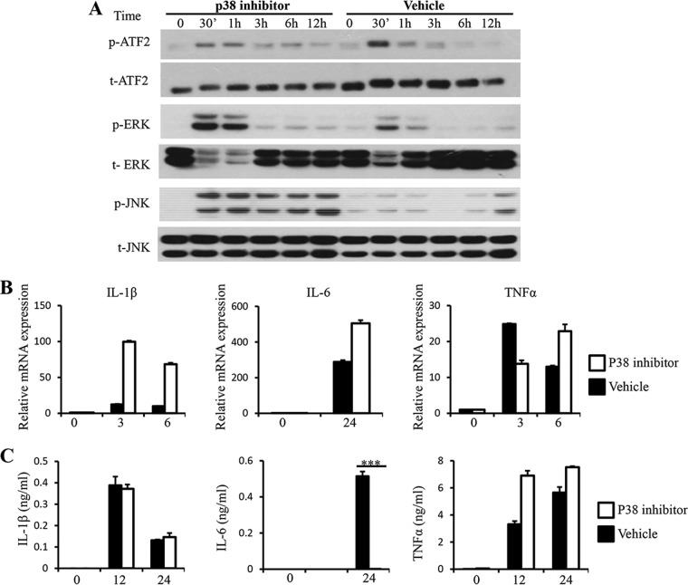
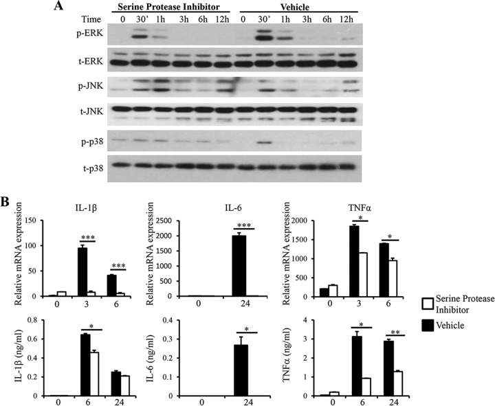
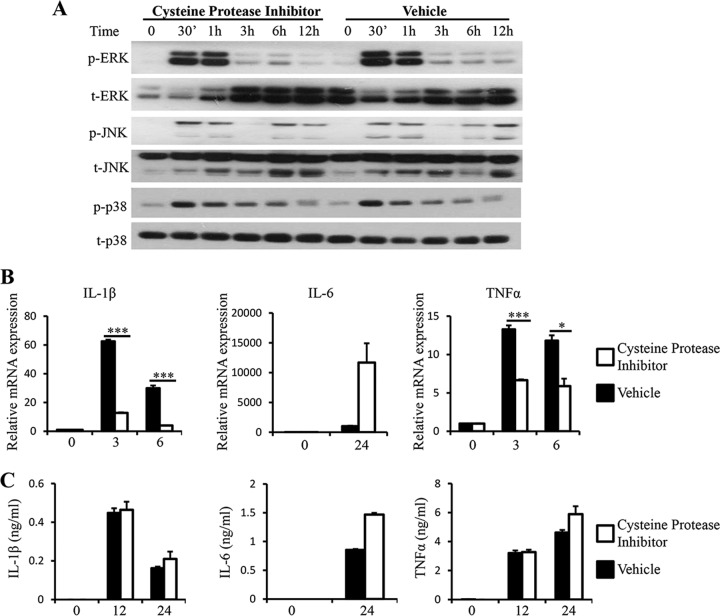
Blastocystis is a common enteric protistan parasite that can cause acute, as well as chronic, infection and is associated with irritable bowel syndrome (IBS). However, the pathogenic status of Blastocystis infection remains unclear. In this study, we found that Blastocystis antigens induced abundant expression of proinflammatory cytokines, including interleukin 1β (IL-1β), IL-6, and tumor necrosis factor alpha (TNF-α), in mouse intestinal explants, in mouse colitis colon, and in macrophages. Further investigation utilizing RAW264.7 murine macrophages showed that Blastocystis treatment in RAW264.7 macrophages induced the activation of ERK, JNK, and p38, the three major groups of mammalian mitogen-activated protein (MAP) kinases that play essential roles in the expression of proinflammatory cytokines. ERK inhibition in macrophages significantly suppressed both mRNA and protein expression of IL-6 and TNF-α and mRNA expression of IL-1β. On the other hand, JNK inhibition resulted in reductions in both c-Jun and ERK activation and significant suppression of all three proinflammatory cytokines at both the mRNA and protein levels. Inhibition of p38 suppressed only IL-6 protein expression with no effect on the expression of IL-1β and TNF-α. Furthermore, we found that serine proteases produced by Blastocystis play an important role in the induction of ERK activation and proinflammatory cytokine expression by macrophages. Our study thus demonstrated for the first time that Blastocystis could induce the expression of various proinflammatory cytokines via the activation of MAP kinases and that infection with Blastocystis may contribute to the pathogenesis of inflammatory intestinal diseases through the activation of inflammatory pathways in host immune cells, such as macrophages.


Figures

FIG 1
FIG 1
Blastocystis treatment upregulates proinflammatory cytokines in mouse intestinal explants and BMDMs. (A) Murine intestinal explants were isolated from C57/BL6 mice and treated with Blastocystis ST-7 (B) lysate. The culture supernatants were tested using ELISA for protein concentrations of proinflammatory cytokines, IL-1β and TNF-α. (B) Mouse BMDMs were generated and treated with either Blastocystis ST-4 (WR-1) or ST-7 (B) at MOIs of 20 and 10, respectively. The mRNA levels of TNF-α, IL-1β, IL-6, and GAPDH were also measured by qRT-PCR. (C) The concentrations of IL-6, TNF-α, and IL-1β in the culture supernatants of BMDMs were measured by ELISA. Each value represents the mean and standard deviation from at least 3 individual experiments. *, P < 0.05; **, P < 0.01; ***, P < 0.005 (representing a statistically significant difference compared to the negative control). ###, P < 0.005 (representing a statistically significant difference between Blastocystis ST-4 and ST-7).
FIG 2
FIG 2
Blastocystis ST-7 (B) induces stronger proinflammatory cytokine expression and MAPK activation in murine macrophages than ST-4 (WR1). RAW264.7 cells were incubated with lysates of either Blastocystis ST-4 (WR1) or ST-7 (B) at MOIs of 20 and 10, respectively. (A) mRNA levels of TNF-α, IL-1β, IL-6, and GAPDH in macrophages were measured by qRT-PCR. The results are normalized to GAPDH and expressed as the mean fold change relative to the 0-h time point, which is set at 1. (B) The concentrations of IL-6, TNF-α, and IL-1β in the culture supernatants of RAW264.7 cells were measured by ELISA. Each value represents the mean and standard deviation from at least 3 individual experiments. *, P < 0.05; **, P < 0.01; ***, P < 0.005 (representing a statistically significant difference compared to the negative control). #, P < 0.05; ##, P < 0.01; ###, P < 0.005 (representing a statistically significant difference between Blastocystis ST-4 and ST-7). (C) S. boulardii lysate does not induce inflammatory cytokine expression in macrophages. RAW264.7 cells were incubated with lysate of either Blastocystis ST-7 (B) (MOI, 10) or S. boulardii (MOI, 20). The mRNA levels of TNF-α, IL-1β, IL-6, and GAPDH in macrophages were measured by qRT-PCR. (D) Representative Western blots of MAPK activation in RAW264.7 cells in response to ST-4 (WR1) or ST-7 (B) lysate stimulation. Cells were lysed and were processed by SDS-PAGE. Membranes were then blotted with phospho-ERK (p-ERK), total ERK (t-ERK), phospho-JNK, total JNK, phospho-p38, and total p38 antibodies. Each blot is representative of three separate experiments.
FIG 3
FIG 3
Inhibition of ERK leads to reduced cytokine expression in response to Blastocystis. RAW264.7 cells were pretreated with either 100 μM PD98059 or ethanol vehicle dissolved in RPMI complete medium for 1 hour prior to stimulation with Blastocysis ST-7 at an MOI of 10 for the indicated times. (A) Representative Western blots for MAPK activation in RAW264.7 cells. The cells were lysed and processed by SDS-PAGE. Membranes were then blotted with phospho-ERK, ERK, phospho-JNK, JNK, phospho-p38, and p38 antibodies. Each blot is representative of three separate experiments. (B) mRNA levels of TNF-α, IL-1β, IL-6, and GAPDH were measured by qRT-PCR. The values are expressed as mean relative transcript levels normalized to GAPDH and are expressed relative to the 0-h time point, which is set at 1. (C) Concentrations of IL-6, TNF-α, and IL-1β in the culture supernatants were measured by ELISA. Each value represents the mean and standard deviation from at least 3 individual experiments. *, P < 0.05; **, P < 0.01; ***, P < 0.005 (representing a statistically significant difference between inhibitor and vehicle treatments).
FIG 4
FIG 4
JNK inhibition leads to reduced c-Jun and ERK activation and expression of proinflammatory cytokines upon Blastocystis treatment. RAW264.7 cells were pretreated with either 20 μM SP100625 or DMSO (dimethyl sulfoxide) vehicle dissolved in RPMI complete medium for 1 hour prior to stimulation with Blastocystis ST-7 (B) at an MOI of 10. (A) Representative Western blots for activation of c-Jun and MAPKs in RAW264.7 cells. The cells were lysed and processed by SDS-PAGE. Membranes were then blotted with phospho-ERK, ERK, phospho-JNK, JNK, phospho-cJun, c-Jun, phospho-p38, and p38 antibodies. Each blot is representative of three separate experiments. (B) mRNA levels of TNF-α, IL-1β, IL-6, and GAPDH were measured by qRT-PCR. The values are expressed as mean relative transcript levels normalized to GAPDH and are expressed relative to the 0-h time point, which is set at 1. (C) Concentrations of IL-6, TNF-α, and IL-1β in the supernatants of RAW264.7 cells were measured by ELISA. Each value represents the mean and standard deviation from at least 3 individual experiments. *, P < 0.05; **, P < 0.01; ***, P < 0.005 (representing a statistically significant difference between inhibitor and vehicle treatments).
FIG 5
FIG 5
Inhibition of p38 leads to increased phosphorylation of ERK and JNK and reduced secretion of IL-6. RAW264.7 cells were pretreated with either 20 μM SB203580 or DMSO vehicle dissolved in RPMI complete medium for 1 hour prior to stimulation with Blastocystis ST-7 at an MOI of 10. (A) Representative Western blots for activation of ATF2, ERK, and JNK. Cells were lysed and processed by SDS-PAGE. Membranes were then blotted with phospho-ERK, ERK, phospho-JNK, JNK, phospho-ATF2, and ATF2 antibodies. Each blot is representative of three separate experiments. (B) mRNA levels of TNF-α, IL-1β, IL-6, and GAPDH were measured by qRT-PCR. The values are expressed as mean relative transcript levels normalized to GAPDH and are expressed relative to the 0-h time point, which is set at 1. (C) Concentrations of IL-6, TNF-α and IL-1β in the culture supernatants were measured by ELISA. Each value represents the mean and standard deviation from at least 3 individual experiments. ***, P < 0.005 (representing a statistically significant difference between inhibitor and vehicle treatments).
FIG 6
FIG 6
Serine protease inhibition leads to decreased ERK activation and proinflammatory cytokine expression. ST-7 (B) lysates were pretreated with 500 μM serine protease inhibitor, AEBSF, for 1 hour before coincubation with RAW264.7 cells at 37°C. (A) Representative Western blots for activation of MAPKs. Cells were lysed, and cell extracts were analyzed by Western blotting using phospho-ERK, total ERK, phospho-JNK, total JNK, phospho-p38, and total p38 antibodies. (B) mRNA levels of TNF-α, IL-1β, IL-6, and GAPDH were measured by qRT-PCR. The values are expressed as mean relative transcript levels normalized to GAPDH and are expressed relative to the 0-h time point, which is set at 1. (C) Concentrations of IL-6, TNF-α, and IL-1β in the supernatants were measured by ELISA. Each value represents the mean and standard deviation from at least 3 individual experiments. *, P < 0.05; **, P < 0.01; ***, P < 0.005 (representing a statistically significant difference between inhibitor and vehicle treatments).
FIG 7
FIG 7
Cysteine protease inhibition results in a decrease in mRNA expression of TNF-α and IL-1β but does not have an effect on protein secretion levels for all three proinflammatory cytokines examined. ST-7 (B) lysates were pretreated with 100 μM cysteine protease inhibitor, E-64, for 1 hour before coincubation with RAW264.7 cells at 37°C. (A) Representative Western blots for activation of MAPKs. Cells were lysed, and cell extracts were analyzed by Western blotting using phospho-ERK, total ERK, phospho-JNK, total JNK, phospho-p38, and p38 antibodies. (B) mRNA levels of TNF-α, IL-1β, IL-6, and GAPDH were measured by qRT-PCR. The values are expressed as mean relative transcript levels normalized to GAPDH and are expressed relative to the 0-h time point, which is set at 1. (C) Concentrations of IL-6, TNF-α, and IL-1β in the supernatants were measured by ELISA. Each value represents the mean and standard deviation from at least 3 individual experiments. *, P < 0.05; ***, P < 0.005 (representing a statistically significant difference between inhibitor and vehicle treatments).

Similar articles

Cited by

References

    1. Strober W, Fuss IJ, Blumberg RS. 2002. The immunology of mucosal models of inflammation. Annu. Rev. Immunol. 20:495–549. 10.1146/annurev.immunol.20.100301.064816. - DOI - PubMed
    1. Kaser A, Zeissig S, Blumberg RS. 2010. Inflammatory bowel disease. Annu. Rev. Immunol. 28:573–621. 10.1146/annurev-immunol-030409-101225. - DOI - PMC - PubMed
    1. Tan KS, Mirza H, Teo JD, Wu B, Macary PA. 2010. Current views on the clinical relevance of Blastocystis spp. Curr. Infect. Dis. Rep. 12:28–35. 10.1007/s11908-009-0073-8. - DOI - PubMed
    1. Tan KS. 2008. New insights on classification, identification, and clinical relevance of Blastocystis spp. Clin. Microbiol. Rev. 21:639–665. 10.1128/CMR.00022-08. - DOI - PMC - PubMed
    1. Clark CG. 1997. Extensive genetic diversity in Blastocystis hominis. Mol. Biochem. Parasitol. 87:79–83. 10.1016/S0166-6851(97)00046-7. - DOI - PubMed

Publication types

MeSH terms

Substances

LinkOut - more resources