. 2014 Nov;351(2):359-72.
doi: 10.1124/jpet.114.217299. Epub 2014 Aug 25.

The limitations of diazepam as a treatment for nerve agent-induced seizures and neuropathology in rats: comparison with UBP302

Affiliations

The limitations of diazepam as a treatment for nerve agent-induced seizures and neuropathology in rats: comparison with UBP302

James P Apland et al. J Pharmacol Exp Ther. 2014 Nov.

Abstract

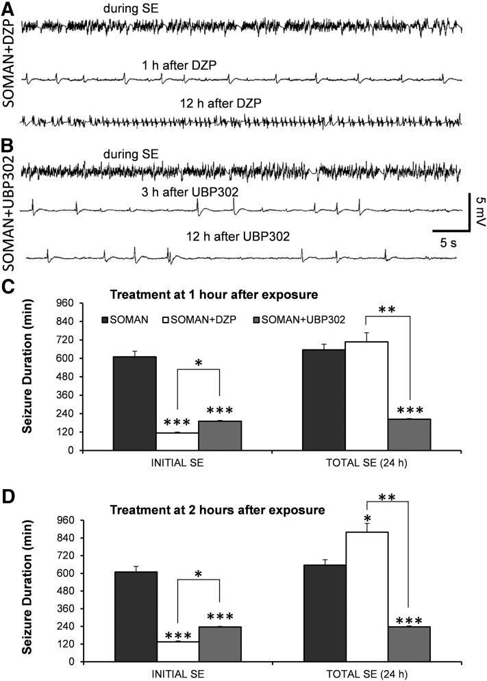
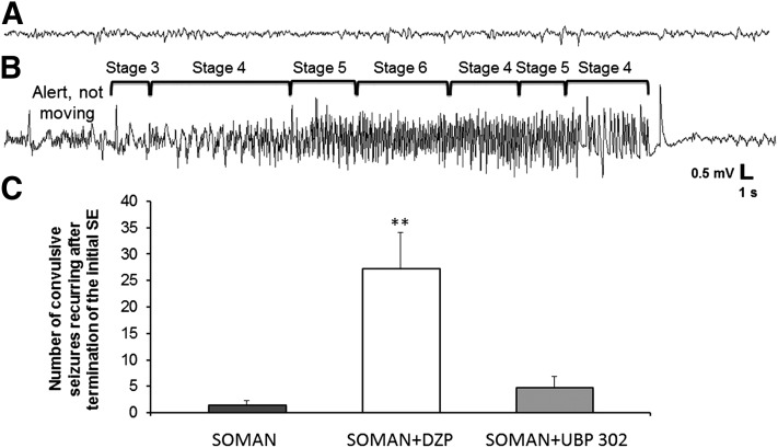
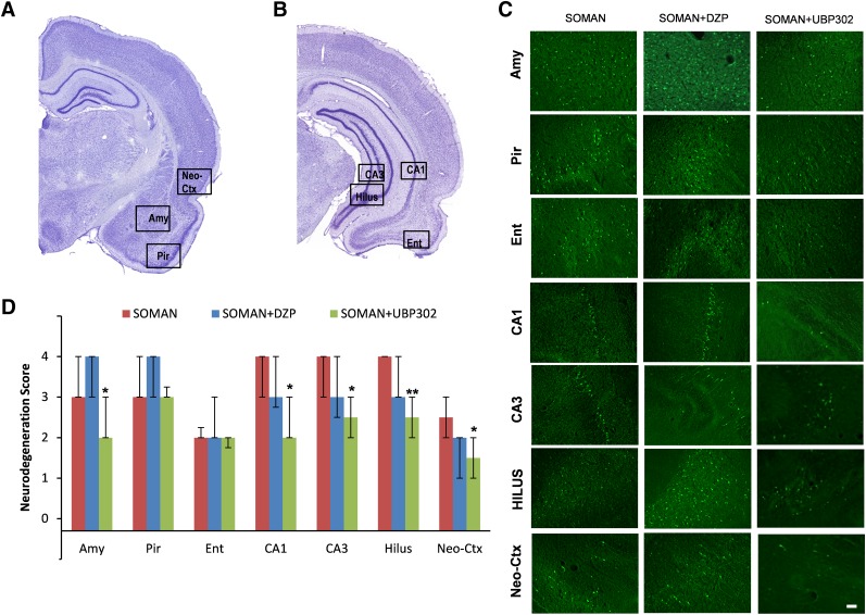
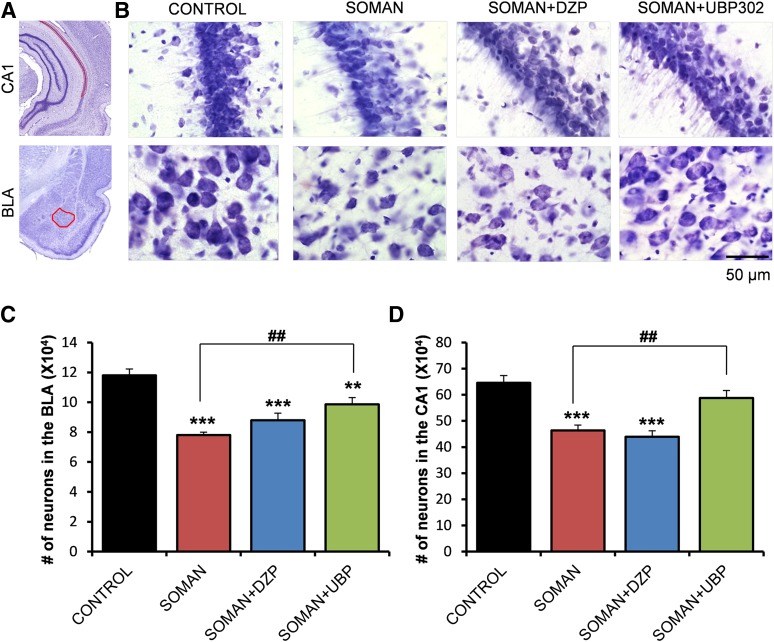
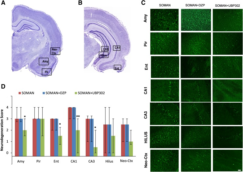
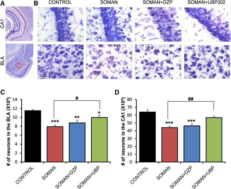
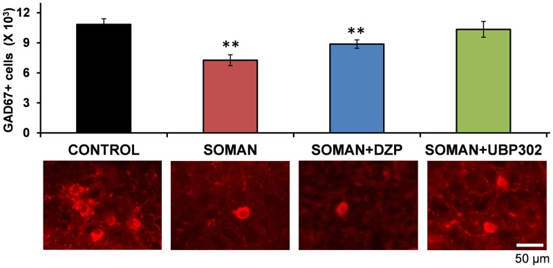
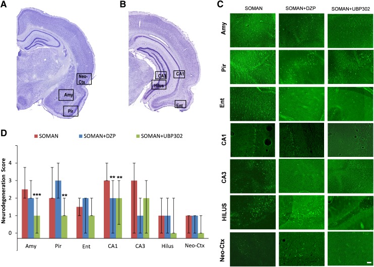
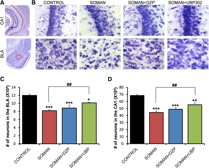
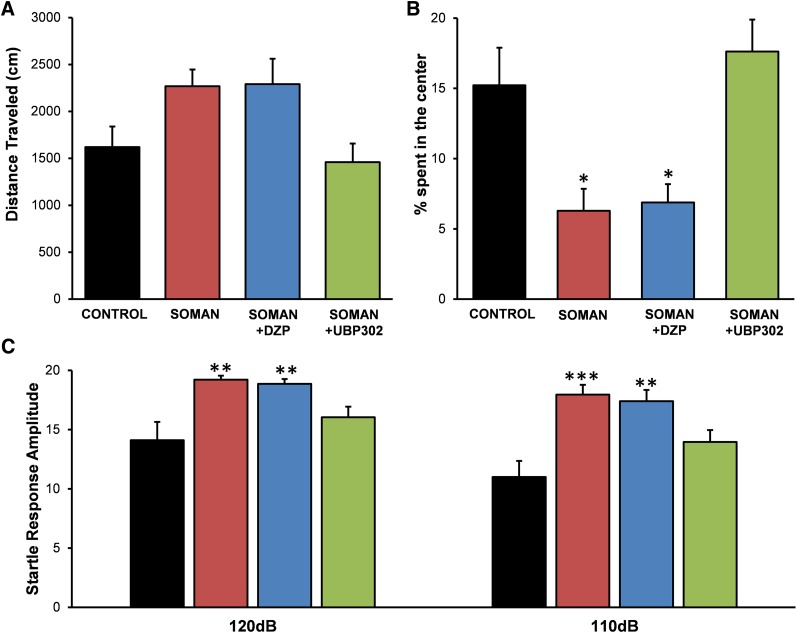
Exposure to nerve agents induces prolonged status epilepticus (SE), causing brain damage or death. Diazepam (DZP) is the current US Food and Drug Administration-approved drug for the cessation of nerve agent-induced SE. Here, we compared the efficacy of DZP with that of UBP302 [(S)-3-(2-carboxybenzyl)willardiine; an antagonist of the kainate receptors that contain the GluK1 subunit] against seizures, neuropathology, and behavioral deficits induced by soman in rats. DZP, administered 1 hour or 2 hours postexposure, terminated the SE, but seizures returned; thus, the total duration of SE within 24 hours after soman exposure was similar to (DZP at 1 hour) or longer than (DZP at 2 hours) that in the soman-exposed rats that did not receive the anticonvulsant. Compared with DZP, UBP302 stopped SE with a slower time course, but dramatically reduced the total duration of SE within 24 hours. Neuropathology and behavior were assessed in the groups that received anticonvulsant treatment 1 hour after exposure. UBP302, but not DZP, reduced neuronal degeneration in a number of brain regions, as well as neuronal loss in the basolateral amygdala and the CA1 hippocampal area, and prevented interneuronal loss in the basolateral amygdala. Anxiety-like behavior was assessed in the open field and by the acoustic startle response 30 days after soman exposure. The results showed that anxiety-like behavior was increased in the DZP-treated group and in the group that did not receive anticonvulsant treatment, but not in the UBP302-treated group. The results argue against the use of DZP for the treatment of nerve agent-induced seizures and brain damage and suggest that targeting GluK1-containing receptors is a more effective approach.


Figures

Fig. 1.
Fig. 1.
DZP terminates soman-induced SE, but does not reduce the total duration of SE within the 24-hour period after soman exposure, as seizures return; UBP302 reduces the total duration of SE within 24 hours. (A and B) Example traces from EEG recordings showing that both DZP and UBP302 (administered 1 hour after soman) terminated the SE induced by soman, but seizure activity returned after DZP administration. (C) Duration of initial SE and total duration of SE within 24 hours after soman exposure, when DZP and UBP302 were administered at 1 hour after soman injection. The three bars on the left show the duration of the initial SE (the SE that started 5–15 minutes after soman exposure and was terminated by DZP or UBP302, or spontaneously in the soman group), whereas the three bars on the right show the total duration of SE (soman: n = 4; soman + DZP: n = 6; soman + UBP302: n = 8). (D) Duration of initial SE and total duration of SE within 24 hours after soman exposure, when DZP and UBP302 were administered at 2 hours after soman injection. The three bars on the left show the duration of the initial SE, whereas the three bars to the right show the total duration of SE (soman: n = 4; soman + DZP: n = 4; soman + UBP302: n = 4). *P < 0.05; **P < 0.01; ***P < 0.001, compared with the soman group (analysis of variance [ANOVA] followed by Bonferroni post hoc test for the initial SE and ANOVA followed by the Games–Howell post hoc test for total SE). *P < 0.05; **P < 0.01 for the comparisons between the DZP-treated and the UBP302-treated groups (ANOVA followed by Fisher’s least significant difference test).
Fig. 2.
Fig. 2.
The number of convulsive seizures that recurred in the DZP-treated rats after termination of the initial SE was greater than in the UBP302-treated rats. (A) EEG baseline before soman exposure. (B) Representative recording of a convulsive seizure recurring after termination of the initial SE by DZP, and its correspondence with the behavioral seizure observations. (C) Number of convulsive seizures that occurred after cessation of the initial SE, within the remaining time of the 24-hour period after soman exposure (soman: n = 4; soman + DZP: n = 6; soman + UBP302: n = 8). **P < 0.01, significantly higher compared with the soman + UBP302 group and the soman group (analysis of variance followed by Holm–Sidak post hoc test).
Fig. 3.
Fig. 3.
UBP302, but not DZP, administered 1 hour after soman exposure, reduced neuronal degeneration in the amygdala, hippocampus, and neocortex 1 day after the exposure. (A and B) Panoramic photomicrographs of Nissl-stained sections showing the brain regions evaluated by FJC staining. (C) Representative photomicrographs of FJC-stained sections from the brain regions where neuronal degeneration was evaluated, for the soman, soman + DZP, and soman + UBP302 groups. (D) Neuropathology scores (median and IQR) for the soman, soman + DZP, and soman + UBP groups (n = 6 for each group) for the amygdala (Amy), piriform cortex (Pir), entorhinal cortex (Ent), CA1, CA3, and hilar areas of the ventral hippocampus (HILUS), and neocortex (Neo-Ctx). *P < 0.05; **P < 0.01, compared with the soman group (Mann–Whitney U test). Original magnification, 100×. Bar, 50 μm.
Fig. 4.
Fig. 4.
UBP302, but not DZP, administered 1 hour after soman exposure, reduced neuronal loss in the BLA and the CA1 hippocampal area, 1 day after the exposure. (A) Panoramic photomicrographs of Nissl-stained half hemispheres outlining the amygdalar nucleus and the hippocampal subfield where stereological analysis was performed. (B) Representative photomicrographs of Nissl-stained sections showing BLA and CA1 cells from the control, soman, soman + DZP, and soman + UBP302 groups. (C and D) Group data (mean and S.E.; n = 6 for each group) of stereological estimation of the total number of Nissl-stained neurons in the BLA (left) and CA1 area (right). **P < 0.01; ***P < 0.001 in comparison with control; ##P < 0.01 in comparison with the soman group (analysis of variance, Dunnett post hoc test). Original magnification, 630×. Bar, 50 μm.
Fig. 5.
Fig. 5.
UBP302, but not DZP, administered 1 hour after soman exposure, reduced neuronal degeneration in the amygdala, CA1, and CA3 dorsal hippocampal areas, and entorhinal cortex, 7 days after the exposure. (A and B) Panoramic photomicrographs of Nissl-stained sections showing the brain regions evaluated by FJC staining. (C) Representative photomicrographs of FJC-stained sections from the brain regions where neuronal degeneration was evaluated, for the soman, soman + DZP, and soman + UBP302 groups. (D) Neuropathology scores (median and IQR) for the soman, soman + DZP, and soman + UBP groups (n = 6 for each group), for the amygdala (Amy), piriform cortex (Pir), entorhinal cortex (Ent), CA1, CA3, and hilar areas of the ventral hippocampus (HILUS), and neocortex (Neo-Ctx). *P < 0.05; **P < 0.01, compared with the soman group (Mann–Whitney U test). Original magnification, 100×. Bar, 50 μm.
Fig. 6.
Fig. 6.
UBP302, but not DZP, administered 1 hour after soman exposure, reduced neuronal loss in the BLA and the CA1 hippocampal area, 7 days after the exposure. (A) Panoramic photomicrographs of Nissl-stained half hemispheres outlining the amygdalar nucleus and the hippocampal subfield where stereological analysis was performed. (B) Representative photomicrographs of Nissl-stained sections showing BLA and CA1 cells from the control, soman, soman + DZP, and soman + UBP302 groups. (C and D) Group data (mean and standard error; n = 6 for each group) of stereological estimation of the total number of Nissl-stained neurons in the BLA (left) and CA1 area (right). *P < 0.05; **P < 0.01; ***P < 0.001, compared with the control; #P < 0.05; ##P < 0.01, compared with the soman group (analysis of variance, Dunnett post hoc test). Original magnification, 630×. Bar, 50 μm.
Fig. 7.
Fig. 7.
UBP302, but not DZP, administered 1 hour after soman exposure, prevented GABAergic interneuronal loss in the BLA, 7 days after the exposure. Group data (mean and standard error; n = 6 for the each group) of stereological estimation of the total number of GAD-67+ neurons in the BLA for the control, soman, soman + DZP, and soman + UBP302 groups. Representative photomicrographs of GAD-67+ interneurons are shown in the lower panel. **P < 0.01, compared with the control (analysis of variance, Dunnett post hoc test). Original magnification, 630×. Bar, 50 μm.
Fig. 8.
Fig. 8.
UBP302, administered 1 hour after soman exposure, reduces neuronal degeneration in the amygdala, piriform cortex, and CA1 hippocampal area, whereas DZP reduces neurodegeneration in the CA1 area, 30 days after the exposure. (A and B) Panoramic photomicrographs of Nissl-stained sections showing the brain regions evaluated by FJC staining. (C) Representative photomicrographs of FJC-stained sections from the brain regions where neuronal degeneration was evaluated, for the soman, soman + DZP, and soman + UBP302 groups. (D) Neuropathology scores (median and IQR) for the soman, soman + DZP, and soman + UBP groups (n = 6 for each group), for the amygdala (Amy), piriform cortex (Pir), entorhinal cortex (Ent), CA1, CA3, and hilar areas of the dorsal hippocampus (HILUS), and neocortex (Neo-Ctx). **P < 0.01; ***P < 0.001, compared with the soman group (Mann–Whitney U test). Original magnification, 100×. Bar, 50 μm.
Fig. 9.
Fig. 9.
UBP302, but not DZP, administered 1 hour after soman exposure, reduced neuronal loss in the BLA and the CA1 hippocampal area, 30 days after the exposure. (A) Panoramic photomicrographs of Nissl-stained half hemispheres outlining the amygdalar nucleus and the hippocampal subfield where stereological analysis was performed. (B) Representative photomicrographs of Nissl-stained sections showing BLA and CA1 cells from the control, soman, soman + DZP, and soman + UBP302 groups. (C and D) Group data (mean and standard error; n = 6 for each group) of stereological estimation of the total number of Nissl-stained neurons in the BLA (left) and CA1 area (right). *P < 0.05; **P < 0.01; ***P < 0.001, compared with the control; ##P < 0.01, compared with the soman group (analysis of variance, Dunnett post hoc test). Original magnification, 630×. Bar, 50 μm.
Fig. 10.
Fig. 10.
UBP302, but not DZP, administered 1 hour after soman exposure, protected against the development of anxiety, 30 days after the exposure. (A) Distance traveled (mean ± standard error) in the open field. (B) Percentage of time spent in the center of the open field. (C) Amplitude of startle responses to 120- and 110-db acoustic stimuli (control: n = 8; soman: n = 10; soman + DZP: n = 9; soman + UBP302: n = 12). *P < 0.05; **P < 0.01; ***P < 0.001 (analysis of variance, Dunnett post hoc comparison with control).

References

    1. Apland JP, Aroniadou-Anderjaska V, Braga MF. (2009) Soman induces ictogenesis in the amygdala and interictal activity in the hippocampus that are blocked by a GluR5 kainate receptor antagonist in vitro. Neuroscience 159:380–389 - PMC - PubMed
    1. Apland JP, Aroniadou-Anderjaska V, Figueiredo TH, Green CE, Swezey R, Yang C, Qashu F, Braga MFM. (2013) Efficacy of the GluK1/AMPA receptor antagonist LY293558 against seizures and neuropathology in a soman-exposure model without pretreatment and its pharmacokinetics after intramuscular administration. J Pharmacol Exp Ther 344:133–140 - PMC - PubMed
    1. Apland JP, Figueiredo TH, Qashu F, Aroniadou-Anderjaska V, Souza AP, Braga MF. (2010) Higher susceptibility of the ventral versus the dorsal hippocampus and the posteroventral versus anterodorsal amygdala to soman-induced neuropathology. Neurotoxicology 31:485–492 - PMC - PubMed
    1. Aroniadou-Anderjaska V, Figueiredo TH, Apland JP, Qashu F, Braga MF. (2009) Primary brain targets of nerve agents: the role of the amygdala in comparison to the hippocampus. Neurotoxicology 30:772–776 - PMC - PubMed
    1. Aroniadou-Anderjaska V, Pidoplichko VI, Figueiredo TH, Almeida-Suhett CP, Prager EM, Braga MF. (2012) Presynaptic facilitation of glutamate release in the basolateral amygdala: a mechanism for the anxiogenic and seizurogenic function of GluK1 receptors. Neuroscience 221:157–169 - PMC - PubMed

Publication types

MeSH terms