. 2015 Feb;64(2):353-9.
doi: 10.2337/db14-0582. Epub 2014 Aug 25.

Regulation of AMPK activation by CD36 links fatty acid uptake to β-oxidation

Affiliations

Regulation of AMPK activation by CD36 links fatty acid uptake to β-oxidation

Dmitri Samovski et al. Diabetes. 2015 Feb.

Abstract

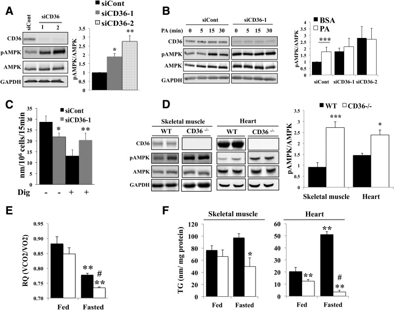
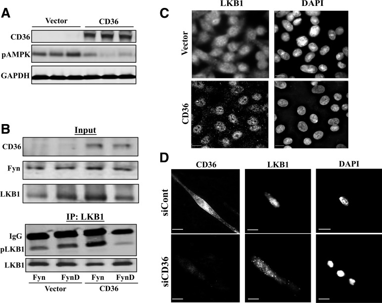
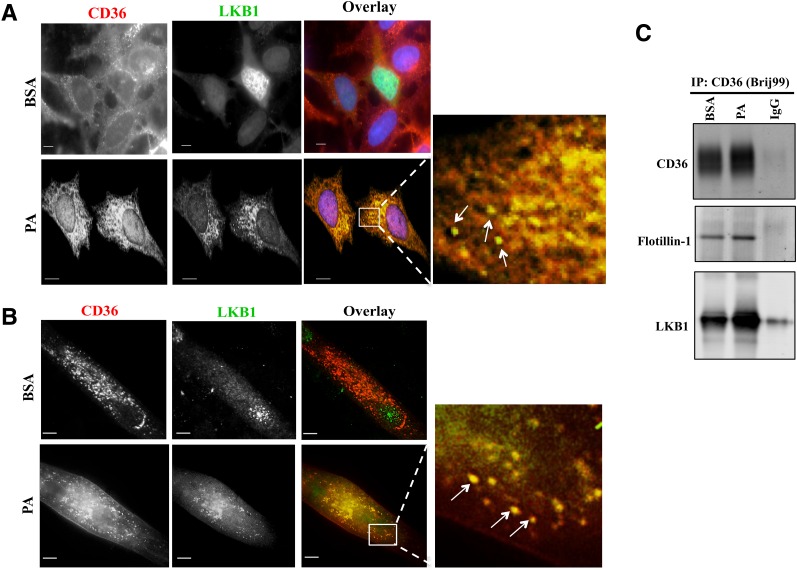
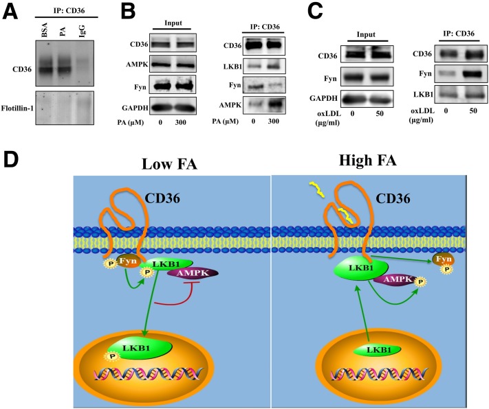
Increases in muscle energy needs activate AMPK and induce sarcolemmal recruitment of the fatty acid (FA) translocase CD36. The resulting rises in FA uptake and FA oxidation are tightly correlated, suggesting coordinated regulation. We explored the possibility that membrane CD36 signaling might influence AMPK activation. We show, using several cell types, including myocytes, that CD36 expression suppresses AMPK, keeping it quiescent, while it mediates AMPK activation by FA. These dual effects reflect the presence of CD36 in a protein complex with the AMPK kinase LKB1 (liver kinase B1) and the src kinase Fyn. This complex promotes Fyn phosphorylation of LKB1 and its nuclear sequestration, hindering LKB1 activation of AMPK. FA interaction with CD36 dissociates Fyn from the protein complex, allowing LKB1 to remain cytosolic and activate AMPK. Consistent with this, CD36(-/-) mice have constitutively active muscle and heart AMPK and enhanced FA oxidation of endogenous triglyceride stores. The molecular mechanism described, whereby CD36 suppresses AMPK, with FA binding to CD36 releasing this suppression, couples AMPK activation to FA availability and would be important for the maintenance of cellular FA homeostasis. Its dysfunction might contribute to the reported association of CD36 variants with metabolic complications of obesity in humans.


Figures

Figure 1
Figure 1
CD36 regulates AMPK phosphorylation and FA oxidation. A: CD36 depletion increases AMPK phosphorylation in myotubes. C2C12 myotubes were transfected with CD36-targeted siRNA (siCD36–1 and siCD36–2) or with a nonspecific siRNA (siCont). Knockdown efficiency relative to siCont: 85% ± 0.74, n = 4, P < 0.01 for siCD36–1; and 66% ± 2.9, n = 4, P < 0.01 for siCD36–2. Cells were serum starved for 16 h in DMEM buffer A containing 5 mmol/L glucose (see Research Design and Methods). Levels of CD36, pAMPK (T172), AMPK, and GAPDH were measured in cell lysates by immunoblotting. Graph shows the quantification by densitometry of pAMPK (T172)/AMPK as a fold change of siCont. Data are reported as the mean ± SE from four experiments. *P < 0.05, **P < 0.01, relative to siCont. B: CD36 is required for FA-induced AMPK activation. C2C12 myotubes treated with siCont or siCD36 were serum starved for 16 h in DMEM buffer A, incubated with 300 μmol/L PA (2:1 with BSA) or BSA (0 min) for the indicated times, and cell lysates were probed for CD36, pAMPK (T172), AMPK, and GAPDH. Graph shows the quantification of pAMPK (T172)/AMPK in cells treated with PA for 15 min as the fold change of levels in siCont. Data are reported as the mean ± SE from three experiments. ***P < 0.001, relative to siCont. C: CD36 depletion increases FA oxidation. C2C12 myotubes were starved for 16 h and processed for FA oxidation (see Research Design and Methods). Plots show PA-supported oxygen consumption (nmol/106 cells/15 min) for intact or Dig-permeabilized myotubes (siCont or CD36 depleted). Data are reported as the mean ± SE from five experiments. **P < 0.01, *P < 0.05 relative to the corresponding siCont. D: CD36 deletion increases AMPK phosphorylation in vivo. Skeletal muscle (gastrocnemius) and hearts, isolated from WT or CD36−/− mice fasted for 18 h were lysed and probed for CD36, pAMPK (T172), AMPK, and GAPDH for skeletal muscle or β-actin for heart (label omitted for clarity). Graph shows the quantification of pAMPK/AMPK levels in gastrocnemius muscle (n = 6 mice) and heart (n = 4 mice). Data are reported as the mean ± SE. ***P < 0.001, *P < 0.05 relative to WT mice. E: CD36 deletion increases FA oxidation in vivo. RQs were measured by indirect calorimetry for WT and CD36−/− mice under ad libitum feeding (18 h) and then during 18 h of fasting. Averaged RQs of fed or fasted WT and CD36−/− mice are shown. Data are reported as the mean ± SE. Significance reflects comparisons to fed WT mice (**P < 0.01) or fasted WT mice (#P < 0.01; n = 6 per group). F: Endogenous TG stores are depleted in CD36-deficient muscle. Left panel: Quadriceps TG content in WT and CD36−/− mice that had been fed or fasted for 18 h. *P < 0.05 compared with fasted WT mice (n = 6 per group). Right panel: Myocardial TG content in WT and CD36−/− mice fed or fasted for 18 h. Comparisons are to fed WT mice (**P < 0.01) or fasted WT mice (#P < 0.01; n = 5 per group). All data are reported as the mean ± SE.
Figure 2
Figure 2
CD36 regulates AMPK activation via Fyn-dependent nuclear sequestration of LKB1. A: Suppression of AMPK phosphorylation by CD36 expression. CHO cells lacking CD36 (Vector) or stably expressing CD36 (CHO-CD36) were serum starved (for 16 h in DMEM buffer A), lysed, and probed (in triplicate) for CD36, pAMPK (T172), and GAPDH. Data are representative of two experiments. B: Fyn phosphorylation of LKB1 is enhanced by CD36 expression. Control (Vector) and CD36-expressing cells (CD36) were transiently transfected with Fyn or FynD; and serum-starved and cell lysates (Input) were probed for CD36, LKB1, and Fyn. LKB1 was immunoprecipitated from cell lysates using mouse monoclonal antibody for LKB1 (clone 5c10; Millipore). LKB1 immunoprecipitates (IP:LKB1) were probed with phosphotyrosine PY100 (pLKB1) and LKB1 antibodies. C: CD36 expression induces nuclear sequestration of LKB1 in CHO cells. CHO cells stably expressing CD36 or Vector controls were serum starved, fixed, and processed for IF as described in Research Design and Methods. The cells were stained with mouse monoclonal LKB1 antibody (clone 5c10; Millipore) and with DAPI to visualize the nuclei. Images are representative of multiple fields from three experiments. Scale bar, 10 μm. D: CD36 depletion in C2C12 myotubes abolishes nuclear sequestration of LKB1. C2C12 myotubes treated with siCD36 or siCont were serum starved for 16 h in DMEM buffer A. LKB1 and CD36 were detected using mouse monoclonal anti-LKB1 (5c10; Millipore) and rat monoclonal anti-CD36 antibodies (MF3; AbD Serotec). Images are representative of multiple fields from three experiments. Scale bar, 10 μm.
Figure 3
Figure 3
PA induces CD36 association with LKB1. A and B: PA addition relocates LKB1 to cytosolic CD36-positive vesicular structures. Micrographs showing PA-induced redistribution of LKB1 and its colocalization with CD36 in CHO-CD36 cells (A) and in myotubes (B). The cells were serum starved, incubated with 300 μmol/L PA (2:1 BSA) or BSA for 15 min, and stained with anti-CD36 (FA6-152 [Abcam] for CHO; MF3 [AbD Serotec] for myotubes) and anti-LKB1 (5c10; Millipore) antibodies. Images are representative of multiple fields from three experiments. Arrows point to overlap between CD36 and LKB1 within cytoplasmic vesicular structures. Scale bar, 10 μm. C: PA addition results in a CD36-LKB1 association within lipid domains. CHO-CD36 cells, serum starved then incubated with 300 μmol/L PA (2:1 BSA) or BSA for 15 min were lysed with buffer containing 1% Brij99. Equal protein amounts were immunoprecipitated (IP:CD36) using anti-CD36 antibody (AF1955; R&D Systems) and nonimmune IgG as a negative control. Immunoprecipitates were resolved by SDS-PAGE, and probed for CD36, flotillin-1, and LKB1. Data are representative of two experiments.
Figure 4
Figure 4
FA and oxLDL differentially regulate the association of CD36 with Fyn, LKB1, and AMPK. A and B: PA reduces the CD36 association with Fyn, while enhancing the association with LKB1 and AMPK. CHO-CD36 cells transiently transfected with Fyn were serum starved, incubated for 15 min with 300 μmol/L PA (2:1 with BSA) or BSA, and lysed with buffer containing 60 mmol/L Octyl. A: Equal amounts of protein were immunoprecipitated using anti-CD36 antibody and nonimmune IgG as a negative control. Immunoprecipitates were resolved by SDS-PAGE, and incubated with antibodies for CD36 and flotillin-1. Data are representative of two experiments. B: Immunoblots of cell lysates (input) were resolved by SDS-PAGE, and probed for CD36, pAMPK (T172), AMPK, Fyn, and GAPDH. CD36 immunoprecipitates (IP:CD36) were probed for CD36, Fyn, LKB1, and AMPK. Data are representative of three experiments. C: oxLDL enhances CD36 association with Fyn. CHO-CD36 cells transiently transfected with Fyn were serum starved and incubated for 15 min with 50 μg/mL oxLDL, then were lysed with buffer containing Octyl. Cell lysates (Input) were probed for CD36, pAMPK (T172), AMPK, Fyn, and GAPDH. CD36 immunoprecipitates were probed for CD36, Fyn, LKB1, and AMPK. Data are representative of three experiments. D: Schematic representation of the mechanism for AMPK regulation by CD36 and FA. A ternary protein complex composed of Fyn, LKB1, and AMPK is assembled with CD36. When exogenous FA concentrations are low (left panel), CD36-bound Fyn can access and phosphorylate (P) LKB1, which induces LKB1 nuclear relocation and reduces the amount of cytosolic LKB1 available to activate AMPK. As a result AMPK is kept quiescent. When exogenous FA concentrations rise (right panel), the enhanced FA interaction with CD36 promotes Fyn dissociation from the protein complex, hindering the access of Fyn to phosphorylate CD36-associated LKB1. The ensuing enrichment in cytosolic LKB1 activates AMPK, which enhances FA oxidation by inactivating acetyl-CoA carboxylase. AMPK also induces cell surface CD36 recruitment (3,5). Thus, the CD36-AMPK pathway integrates FA uptake and FA catabolism. When CD36 is depleted, the lack of nuclear sequestration of LKB1 results in constitutively active AMPK that cannot be further activated by FA. The data suggest that CD36 dysfunction impairs AMPK lipid-sensing ability.

References

    1. Hardie DG. AMPK: a target for drugs and natural products with effects on both diabetes and cancer. Diabetes 2013;62:2164–2172 - PMC - PubMed
    1. Shirwany NA, Zou MH. AMPK: a cellular metabolic and redox sensor. A minireview. Front Biosci (Landmark Ed) 2014;19:447–474 - PMC - PubMed
    1. Glatz JF, Luiken JJ, Bonen A. Membrane fatty acid transporters as regulators of lipid metabolism: implications for metabolic disease. Physiol Rev 2010;90:367–417 - PubMed
    1. Ruderman NB, Carling D, Prentki M, Cacicedo JM. AMPK, insulin resistance, and the metabolic syndrome. J Clin Invest 2013;123:2764–2772 - PMC - PubMed
    1. Pepino MY, Kuda O, Samovski D, Abumrad NA. Structure-function of CD36 and importance of fatty acid signal transduction in fat metabolism. Annu Rev Nutr 2014;34:281–303 - PMC - PubMed

Publication types

MeSH terms