. 2014 Sep 9;111(36):13046-51.
doi: 10.1073/pnas.1406050111. Epub 2014 Aug 25.

Unfolded protein response activation reduces secretion and extracellular aggregation of amyloidogenic immunoglobulin light chain

Affiliations

Unfolded protein response activation reduces secretion and extracellular aggregation of amyloidogenic immunoglobulin light chain

Christina B Cooley et al. Proc Natl Acad Sci U S A. .

Abstract

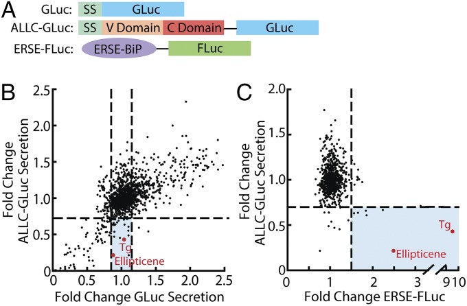
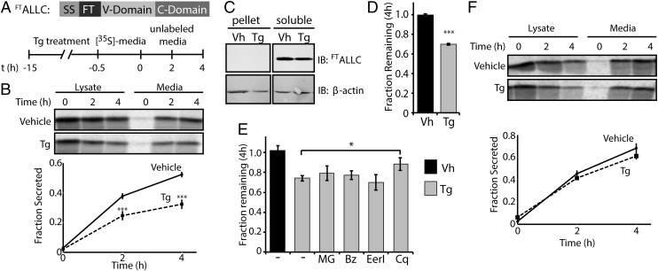
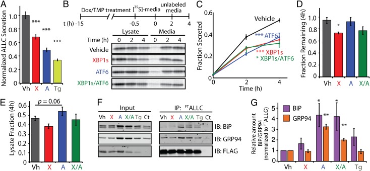
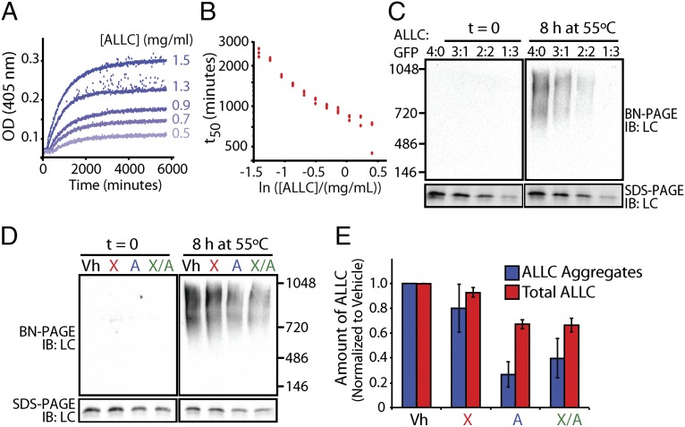
Light-chain amyloidosis (AL) is a degenerative disease characterized by the extracellular aggregation of a destabilized amyloidogenic Ig light chain (LC) secreted from a clonally expanded plasma cell. Current treatments for AL revolve around ablating the cancer plasma cell population using chemotherapy regimens. Unfortunately, this approach is limited to the ∼ 70% of patients who do not exhibit significant organ proteotoxicity and can tolerate chemotherapy. Thus, identifying new therapeutic strategies to alleviate LC organ proteotoxicity should allow AL patients with significant cardiac and/or renal involvement to subsequently tolerate established chemotherapy treatments. Using a small-molecule screening approach, the unfolded protein response (UPR) was identified as a cellular signaling pathway whose activation selectively attenuates secretion of amyloidogenic LC, while not affecting secretion of a nonamyloidogenic LC. Activation of the UPR-associated transcription factors XBP1s and/or ATF6 in the absence of stress recapitulates the selective decrease in amyloidogenic LC secretion by remodeling the endoplasmic reticulum proteostasis network. Stress-independent activation of XBP1s, or especially ATF6, also attenuates extracellular aggregation of amyloidogenic LC into soluble aggregates. Collectively, our results show that stress-independent activation of these adaptive UPR transcription factors offers a therapeutic strategy to reduce proteotoxicity associated with LC aggregation.

Keywords: ER proteostasis; amyloid.


Conflict of interest statement

The authors declare no conflict of interest.

Figures

Fig. 1.
Fig. 1.
ALLC-GLuc reporter screening identifies UPR modulators able to reduce secretion of amyloidogenic LC. (A) Schematic of GLuc, ALLC-GLuc, and ERSE-FLuc reporter constructs used in stable ARPE19 or HEK293T-Rex cell lines. ERSE-FLuc transcriptional reporter includes the ERSE-containing portion of the BiP promoter upstream of the firefly luciferase (FLuc) gene. SS, signal sequence. (B) Plot of LOPAC screening data comparing ALLC-GLuc secretion vs. GLuc secretion. ARPE-19 cells stably expressing ALLC-GLuc or GLuc alone were treated with compound (10 μM) for 24 h. Shaded blue section indicates compounds that reduce ALLC-GLuc secretion (>25%) and do not affect GLuc secretion (<±15% from DMSO control). (C) Plot of LOPAC screening results comparing ALLC-GLuc secretion (ARPE-19 cells, 24-h treatment) vs. UPR activation (ERSE-FLuc expressing HEK293T-Rex cells, 18-h treatment). Compounds falling below the horizontal line reduce ALLC-GLuc secretion by 25%. Compounds falling to the right of the vertical line increase ERSE-FLuc expression >1.5-fold relative to DMSO control. Compounds depicted in red are known activators of the UPR.
Fig. 2.
Fig. 2.
Thapsigargin selectively reduces the secretion of amyloidogenic LC and induces its degradation. (A) Schematic of the FTALLC construct used in pulse-chase experiments and the metabolic labeling protocol used. [35S]-labeled FTALLC was immunopurified from media and lysates collected from transfected HEK293T-Rex cells following a 15-h treatment with thapsigargin (Tg, 500 nM). (B) Representative autoradiogram and quantification of [35S]-labeled FTALLC in pulse-chase experiment described in A. Fraction secreted was calculated as described in Materials and Methods (18) (n ≥ 3). (C) Immunoblots measuring soluble and insoluble (pellet) levels of FTALLC after 15-h pretreatment with Tg. (D) Graph depicting total [35S]-labeled FTALLC (combined media and lysate protein levels as in B) remaining at 4 h in HEK293T-Rex cells following a 15-h pretreatment with 500 nM Tg (n ≥ 3). The fraction remaining was calculated as described in Materials and Methods (18). (E) Graph depicting the fraction recovery of total [35S]- labeled FTALLC at 4 h in HEK293T-Rex cells incubated in the presence of thapsigargin (Tg; 500 nM, 15 h) and the proteasome inhibitors bortezomib (Bz; 20 µM) or MG132 (MG; 20 µM), the ERAD inhibitor eeyarestatin I (EerI; 20 µM), or the autophagy inhibitor chloroquine (Cq; 100 µM). Inhibitors were incubated for 4 h before [35S] metabolic labeling and included throughout the labeling protocol shown in A. Fraction remaining was calculated as in D (n ≥ 2). (F) Representative autoradiogram and quantification of [35S]-labeled FTJTO immunopurified from media and lysates collected from transfected HEK293T-Rex cells following the same protocol and quantification as in A and B (n ≥ 3). *P < 0.05; ***P < 0.005. All error bars represent the SEM from biological replicates.
Fig. 3.
Fig. 3.
Stress-independent activation of XBP1s and/or ATF6 decreases the secretion of amyloidogenic LC. (A) ALLC levels determined by ELISA in conditioned media from HEK293DAX cells (40) expressing FTALLC following a 15-h preactivation of XBP1s (X; Dox, 1 μg/mL) or ATF6 (A; TMP, 10 μM) or by thapsigargin-induced stress (Tg, 500 nM). ALLC secretion levels were normalized to vehicle (Vh; DMSO) conditions (n = 3). (B) Representative autoradiogram of [35S]-labeled FTALLC immunopurified from media and lysates collected from transfected HEK293T-Rex cells following a 15-h preactivation of XBP1s (X; Dox, 1 μg/mL), ATF6 (A; TMP, 10 μM), or both (X/A). The metabolic labeling protocol used is shown. (C) Quantification of fraction secreted from autoradiograms as shown in B (n ≥ 3). (D) Graph depicting the total [35S]-labeled FTALLC remaining at 4 h in lysate and media following a 15-h preactivation of XBP1s, ATF6, or both in HEK293DAX (n ≥ 3). (E) Graph depicting the total intracellular [35S]-labeled FTALLC in lysates at 4 h in HEK293DAX cells following a 15-h preactivation of XBP1s, ATF6, or both (n ≥ 3). The lysate fraction was calculated by dividing the lysate [35S]-labeled FTALLC signal at 4 h by the total [35S]-labeled FTALLC at t = 0. (F) Immunoblot showing the recovery of the ER chaperones BiP and GRP94 in FLAG immunopurifications from cross-linked HEK293DAX cells expressing FTALLC following 15-h pretreatment with thapsigargin (Tg), XBP1s (X), ATF6 (A), or XBP1s and ATF6 (X/A), as in B. HEK293DAX cells expressing untagged ALLC is shown as a control (Ct). (G) Quantification of F achieved by comparing the signal under various conditions to vehicle, and by normalizing to the recovered FTALLC. *P < 0.05; **P < 0.01; ***P < 0.005. All error bars represent the SEM from biological replicates.
Fig. 4.
Fig. 4.
Stress-independent activation of XBP1s and especially ATF6 reduces aggregation of cell-secreted ALLC. (A) Timecourses of recombinant ALLC aggregation at the indicated concentrations. Samples were incubated at 44 °C for the indicated time, and aggregation was measured by turbidity at 405 nm. (B) Plot of the t50 of recombinant ALLC aggregation at the indicated concentration from data as shown in A; n = 3 replicates are shown. (C) Immunoblots for BN-PAGE and SDS-PAGE of media conditioned on HEK293DAX cells expressing FTALLC for 24 h. The media was diluted with media conditioned on GFP-expressing cells, as indicated, and incubated at 55 °C for 8 h. (D) Representative immunoblots for BN-PAGE and SDS-PAGE of media conditioned on HEK293DAX cells following a 16-h preactivation of XBP1s (X), ATF6 (A), or XBP1s and ATF6 (X/A), as in Fig. 3B. FTALLC aggregation was induced by incubating the media for 8 h at 55 °C. (E) Graph depicting the quantification of soluble aggregates and total ALLC from BN-PAGE and SDS-PAGE immunoblots, as shown in D, normalized to vehicle. Error bars represent SEM from biological replicates (n = 3).

References

    1. Cohen AD, Comenzo RL. Systemic light-chain amyloidosis: Advances in diagnosis, prognosis, and therapy. Hematology (Am Soc Hematol Educ Program) 2010;2010:287–294. - PubMed
    1. Buxbaum JN, Linke RP. A molecular history of the amyloidoses. J Mol Biol. 2012;421(2-3):142–159. - PubMed
    1. Jones NF, Hilton PJ, Tighe JR, Hobbs JR. Treatment of “primary” renal amyloidosis with melphalan. Lancet. 1972;2(7778):616–619. - PubMed
    1. Jaccard A, et al. Myélome Autogreffe (MAG) and Intergroupe Francophone du Myélome (IFM) Intergroup High-dose melphalan versus melphalan plus dexamethasone for AL amyloidosis. N Engl J Med. 2007;357(11):1083–1093. - PubMed
    1. Kastritis E, et al. Bortezomib with or without dexamethasone in primary systemic (light chain) amyloidosis. J Clin Oncol. 2010;28(6):1031–1037. - PubMed

Publication types

MeSH terms

LinkOut - more resources