

Small bowel obstruction following computed tomography and magnetic resonance enterography using psyllium seed husk as an oral contrast agent
- PMID: 25157531
- PMCID: PMC4144458
- DOI: 10.1155/2014/501043
Small bowel obstruction following computed tomography and magnetic resonance enterography using psyllium seed husk as an oral contrast agent
Abstract
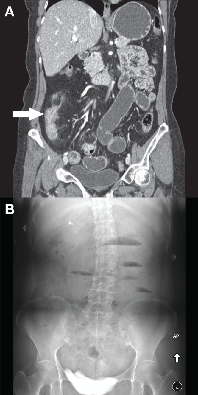
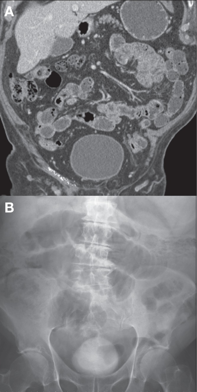
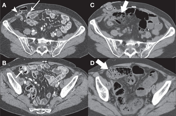
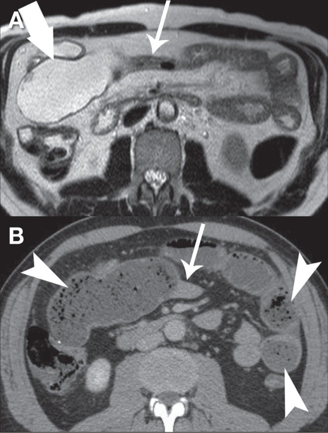
The authors report a case series describing four patients who developed small bowel obstruction following the use of psyllium seed husk as an oral contrast agent for computed tomography or magnetic resonance enterography. Radiologists who oversee computed tomography and magnetic resonance enterography should be aware of this potential complication when using psyllium seed husk and other bulking agents, particularly when imaging patients with known or suspected small bowel strictures or active inflammation.
Les auteurs rendent compte d’une série de cas de quatre patients qui ont présenté une obstruction du grêle après avoir consommé l’enveloppe de psyllium comme agent oral de contraste en vue d’une tomodensitométrie ou d’une entérographie par résonance magnétique. Les radiologues qui supervisent les tomodensitométries et les entérographies par résonance magnétique devraient connaître cette complication potentielle lorsqu’ils utilisent l’enveloppe de psyllium et les autres agents gonflants, notamment lorsque les patients qui se soumettent à l’imagerie présentent un rétrécissement connu ou présumé ou une inflammation active du grêle.
Figures
References
-
- Bernstein CN, Greenberg H, Boult I, Chubey S, Leblanc C, Ryner L. A prospective comparison study of MRI versus small bowel follow-through in recurrent Crohn’s disease. Am J Gastroenterol. 2005;100:2493–502. - PubMed
-
- Hara AK, Leighton JA, Heigh RI, et al. Crohn disease of the small bowel: Preliminary comparison among CT enterography, capsule endoscopy, small-bowel follow-through, and ileoscopy. Radiology. 2006;238:128–34. - PubMed
-
- Lee SS, Kim AY, Yang SK, et al. Crohn disease of the small bowel: Comparison of CT enterography, MR enterography, and small-bowel follow-through as diagnostic techniques. Radiology. 2009;251:751–61. - PubMed
-
- Frokjaer JB, Larsen E, Steffensen E, Nielsen AH, Drewes AM. Magnetic resonance imaging of the small bowel in Crohn’s disease. Scand J Gastroenterol. 2005;40:832–42. - PubMed
MeSH terms
Substances
LinkOut - more resources
Full Text Sources
Other Literature Sources
Medical
