. 2014 Oct;124(10):4240-56.
doi: 10.1172/JCI74072. Epub 2014 Aug 26.

SORCS1 is necessary for normal insulin secretory granule biogenesis in metabolically stressed β cells

SORCS1 is necessary for normal insulin secretory granule biogenesis in metabolically stressed β cells

Melkam A Kebede et al. J Clin Invest. 2014 Oct.

Abstract

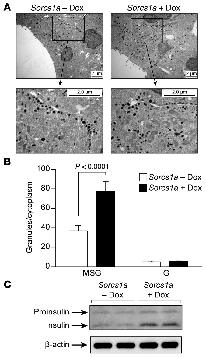
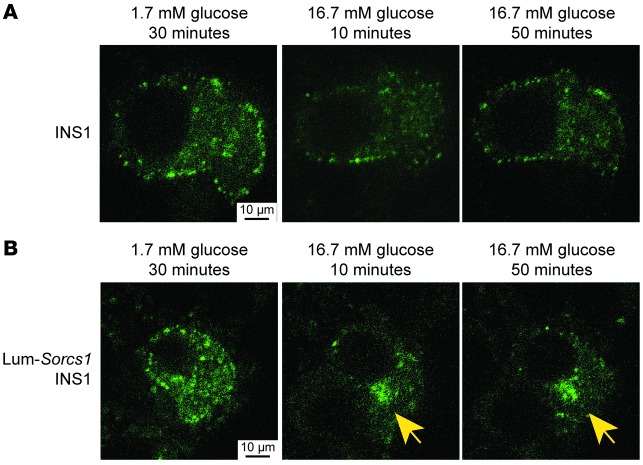
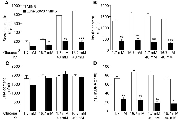
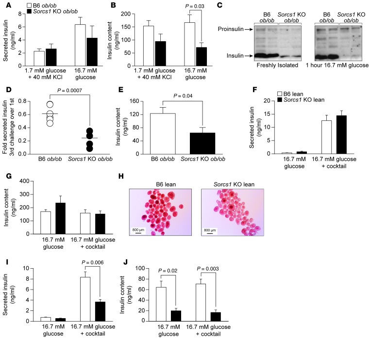
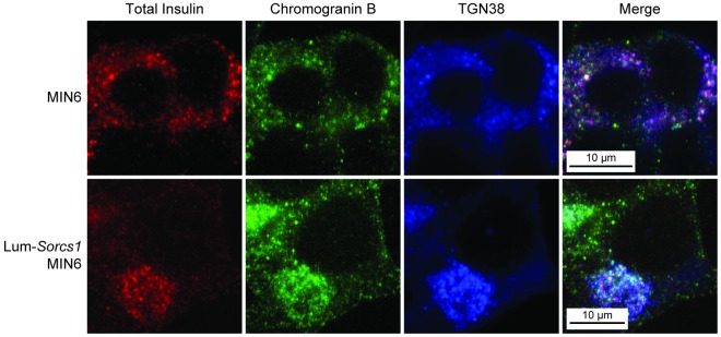
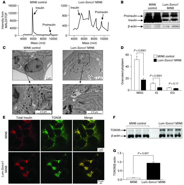
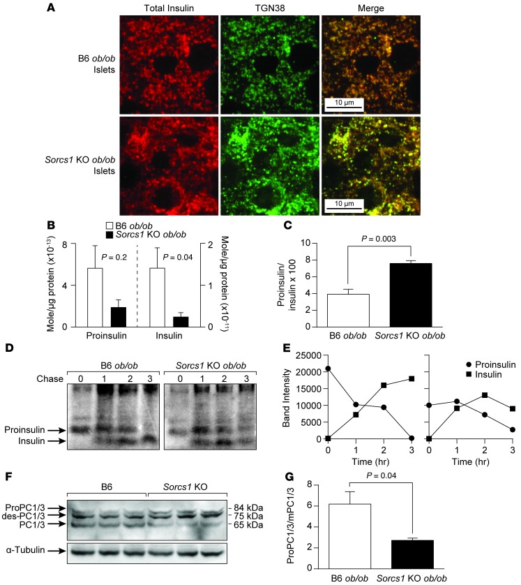
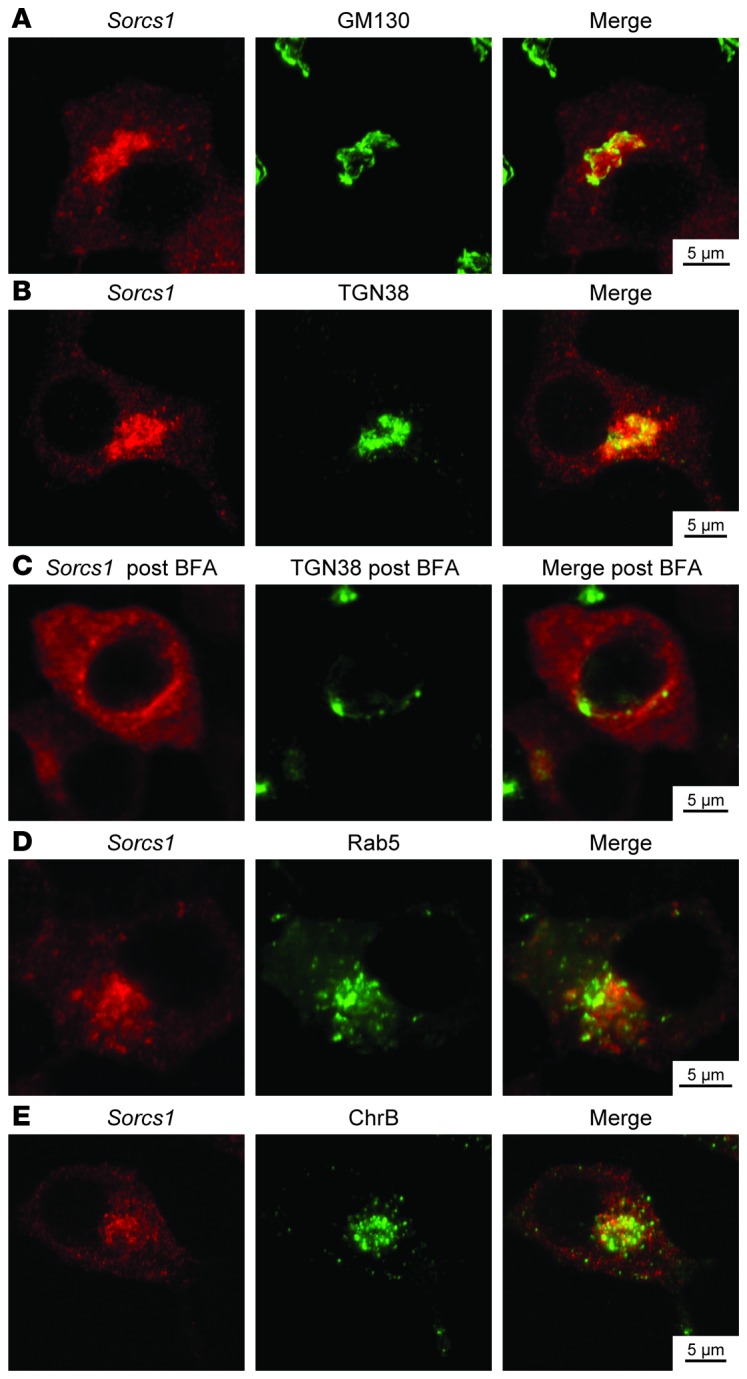
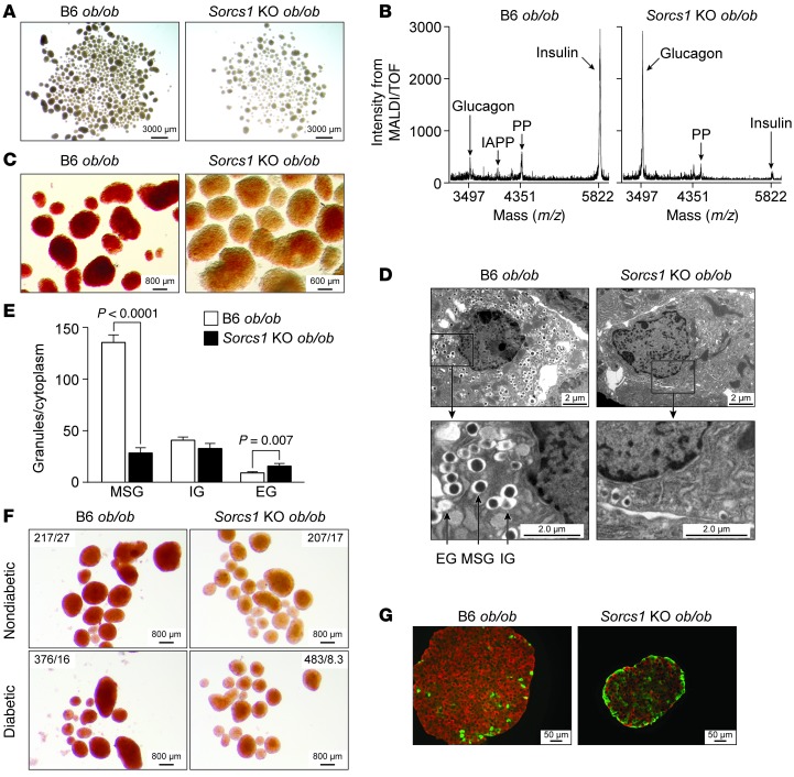
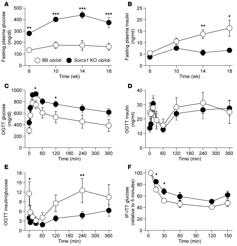
We previously positionally cloned Sorcs1 as a diabetes quantitative trait locus. Sorcs1 belongs to the Vacuolar protein sorting-10 (Vps10) gene family. In yeast, Vps10 transports enzymes from the trans-Golgi network (TGN) to the vacuole. Whole-body Sorcs1 KO mice, when made obese with the leptin(ob) mutation (ob/ob), developed diabetes. β Cells from these mice had a severe deficiency of secretory granules (SGs) and insulin. Interestingly, a single secretagogue challenge failed to consistently elicit an insulin secretory dysfunction. However, multiple challenges of the Sorcs1 KO ob/ob islets consistently revealed an insulin secretion defect. The luminal domain of SORCS1 (Lum-Sorcs1), when expressed in a β cell line, acted as a dominant-negative, leading to SG and insulin deficiency. Using syncollin-dsRed5TIMER adenovirus, we found that the loss of Sorcs1 function greatly impairs the rapid replenishment of SGs following secretagogue challenge. Chronic exposure of islets from lean Sorcs1 KO mice to high glucose and palmitate depleted insulin content and evoked an insulin secretion defect. Thus, in metabolically stressed mice, Sorcs1 is important for SG replenishment, and under chronic challenge by insulin secretagogues, loss of Sorcs1 leads to diabetes. Overexpression of full-length SORCS1 led to a 2-fold increase in SG content, suggesting that SORCS1 is sufficient to promote SG biogenesis.


Figures

Figure 10
Figure 10. Overexpression of full-length Sorcs1a in INS 832/13 cells increases MSGs and insulin content.
(A) β Cell ultrastructure of INS 832/13 cells before and 18 hours after induction with 25 ng/ml doxycycline (Dox). (B) Quantitation of SGs per β cell cytoplasm. (C) Immunoblot analysis of total insulin in lysate from INS 832/13 cells before and 18 hours after induction with 25 ng/ml doxycycline. Data are represented as mean ± SEM. IG, immature granules; MSG, mature secretory granules.
Figure 9
Figure 9. Lum-Sorcs1–expressing cells have a defect in mature secretory granule replenishment.
Confocal microscopy of control (A) and INS1 832/13 cells transiently transfected with luminal Sorcs1 (B) followed by adenoviral infection with syncollin-dsRed5TIMER. Cells were left to recover for 36–48 hours and analyzed after 30 minutes of exposure to 1.7 mM glucose (left) or 10 minutes (middle) or 50 minutes (right) of exposure to 16.7 mM glucose. Yellow arrows identify perinuclear accumulation of syncollin.
Figure 8
Figure 8. Loss of Sorcs1 action impairs insulin secretion.
Secreted insulin (A), insulin content (B), DNA content (C), and insulin/DNA content (D) in control MIN6 and Lum-Sorcs1 MIN6 cells (n = 3). Data are represented as mean ± SEM (*P < 0.05, **P < 0.005, ***P < 0.0001).
Figure 7
Figure 7. Sorcs1 deletion impairs insulin secretion under metabolic stress.
(A and B) Insulin secretion (A) and insulin content (B) in response to a single challenge of 1.7 mM glucose combined with 40 mM KCl or 16.7 mM glucose in B6 ob/ob (n = 5) or Sorcs1 KO ob/ob (n = 5) islets. (C) Representative immunoblotting of freshly isolated B6 ob/ob and Sorcs1 KO ob/ob islet lysates before (left) and 1 hour after (right) exposure to 16.7 mM glucose. Antibody against total insulin, which recognized both the pro- and the mature form of insulin, was used. (D and E) Insulin secretion (D) and insulin content (E) in B6 ob/ob (n = 5) and Sorcs1 KO ob/ob (n = 4) islets after 3 repeated secretagogue challenges. (F and G) Secreted insulin (F) and insulin content (G) from isolated islets of (n = 4) B6 and Sorcs1 KO lean mice cultured for 24 hours with 16.7 mM glucose. (H) Dithizone staining of B6 and Sorcs1 KO lean islets cultured for 18 hours with 16.7 mM glucose and 0.5 mM palmitate. (I and J) Secreted insulin (I) and insulin content (J) from isolated islets of (n = 4) B6 and Sorcs1 KO lean mice cultured for 24 hours with 16.7 mM glucose and 0.5 mM palmitate. Data are represented as mean ± SEM.
Figure 6
Figure 6. Total insulin is stalled in the TGN in Lum-Sorcs1–expressing MIN6 cells.
Triple staining of total insulin (red), chromogranin B (green), and TGN38 (blue) in control and Lum-Sorcs1–expressing MIN6 cells.
Figure 5
Figure 5. Lum-Sorcs1 acts as a dominant-negative.
(AF) MALDI/TOF analysis (A), immunoblotting of proinsulin and mature insulin (B), β cell ultrastructure (C), quantitation of SGs per β cell cytoplasm (D), double staining of total insulin (red) and TGN38 (green) (E), and representative immunoblotting for TGN38 and β-actin (F) of control and Lum-Sorcs1–expressing MIN6 cells. (G) Quantitation of TGN38 immunoblot as a ratio of TGN38 to β-actin. EG, empty granules; IG, immature granules; MSG, mature secretory granules. Data are represented as mean ± SEM.
Figure 4
Figure 4. Loss of Sorcs1 impairs proinsulin and proPC1/3 processing and increases islet insulin degradation.
(A) Double staining of islet cryosections for total insulin (red) and TGN38 (green) in B6 ob/ob and Sorcs1 KO ob/ob mice. (B and C) Mass spectrometry analysis of proinsulin and mature insulin content (B) and proinsulin-to-insulin ratio (C) in B6 ob/ob and Sorcs1 KO ob/ob islets. (D) Proinsulin processing in B6 ob/ob and Sorcs1 KO ob/ob islets. (E) Densitometric scanner analysis of the proinsulin and insulin bands in D. (F) Representative immunoblotting of freshly isolated B6 and Sorcs1 KO islet lysates probed for total PC1/3 and α-tubulin. (G) Quantitation of PC1/3 immunoblot as a ratio of proPC1/3 and des-PC1/3 to mature PC1/3. Data are represented as mean ± SEM.
Figure 3
Figure 3. Overexpressed Sorcs1a colocalizes with perinuclear Golgi stacks and some post-Golgi compartments.
Double staining with myc-Sorcs1 (red) and either the cis-Golgi matrix protein GM130 (A), the trans-Golgi protein TGN38 before (B) or after (C) brefeldin A treatment, the early endosomal protein Rab5 (D), or the SG marker chromogranin B (E) (green) of INS1 832/13 cells stably transfected with myc-tagged inducible Sorcs1a plasmid 18 hours after induction with 25 ng/ml doxycycline.
Figure 2
Figure 2. Deletion of Sorcs1 leads to severe depletion of islet insulin content and mature secretory granules (SGs).
(AD) Brightfield illumination (A), MALDI/TOF spectra (B), dithizone staining (C), and β cell ultrastructure (D) of isolated B6 ob/ob and Sorcs1 KO ob/ob islets. (E) Quantification of SGs per β cell cytoplasm from B6 ob/ob and Sorcs1 KO ob/ob β cells. (F) Brightfield images after dithizone staining of islets from nondiabetic and diabetic B6 ob/ob and Sorcs1 KO ob/ob mice. Fasting plasma glucose (mg/dl) and insulin (ng/ml) of the mice are shown as glucose/insulin. (G) Double staining of insulin (red) and glucagon (green) of isolated B6 ob/ob and Sorcs1 KO ob/ob islets. Granule quantification was carried out from electron microscopy images from 3 mice per genotype and 20 β cell fields per mouse. Data are represented as mean ± SEM. EG, empty granules; IAPP, islet-amyloid polypeptide; IG, immature granules; MSG, mature secretory granules; PP, pancreatic polypeptide.
Figure 1
Figure 1. Deletion of Sorcs1 in the leptin-deficient mice leads to diabetes.
(A and B) Plasma glucose (A) and insulin (B) levels following a 4-hour fast in female B6 ob/ob (n = 12) and Sorcs1 KO ob/ob (n = 10) mice as a function of age. (CE) Plasma glucose (C), insulin (D), and insulin-to-glucose ratio (E) during an oral glucose tolerance test (OGTT) in 20-week-old female B6 ob/ob (n = 9) and Sorcs1 KO ob/ob (n = 12) mice. (F) Glucose during an intraperitoneal insulin tolerance test (IP-ITT) in 4-hour-fasted 12-week-old female B6 ob/ob (n = 8) and Sorcs1 KO ob/ob (n = 12) mice. Data are represented as mean ± SEM (*P < 0.05, **P < 0.005, ***P < 0.0001).

References

    1. Groop L, Lyssenko V. Genetic basis of β-cell dysfunction in man. Diabetes Obes Metab. 2009;11(suppl 4):149–158. - PubMed
    1. Steiner DF. The proprotein convertases. Curr Opin Chem Biol. 1998;2(1):31–39. doi: 10.1016/S1367-5931(98)80033-1. - DOI - PubMed
    1. Seidah NG, Prat A. The biology and therapeutic targeting of the proprotein convertases. Nat Rev Drug Discov. 2012;11(5):367–383. doi: 10.1038/nrd3699. - DOI - PubMed
    1. Vindrola O, Lindberg I. Release of the prohormone convertase PC1 from AtT-20 cells. Neuropeptides. 1993;25(2):151–160. doi: 10.1016/0143-4179(93)90096-S. - DOI - PubMed
    1. Benjannet S, Reudelhuber T, Mercure C, Rondeau N, Chretien M, Seidah NG. Proprotein conversion is determined by a multiplicity of factors including convertase processing, substrate specificity, and intracellular environment. Cell type-specific processing of human prorenin by the convertase PC1. J Biol Chem. 1992;267(16):11417–11423. - PubMed

Publication types