. 2014 Oct 15;394(2):197-205.
doi: 10.1016/j.ydbio.2014.08.014. Epub 2014 Aug 23.

Local and global dynamics of the basement membrane during branching morphogenesis require protease activity and actomyosin contractility

Affiliations

Local and global dynamics of the basement membrane during branching morphogenesis require protease activity and actomyosin contractility

Jill S Harunaga et al. Dev Biol. .

Abstract

Many epithelial tissues expand rapidly during embryonic development while remaining surrounded by a basement membrane. Remodeling of the basement membrane is assumed to occur during branching morphogenesis to accommodate epithelial growth, but how such remodeling occurs is not yet clear. We report that the basement membrane is highly dynamic during branching of the salivary gland, exhibiting both local and global remodeling. At the tip of the epithelial end bud, the basement membrane becomes perforated by hundreds of well-defined microscopic holes at regions of rapid expansion. Locally, this results in a distensible, mesh-like basement membrane for controlled epithelial expansion while maintaining tissue integrity. Globally, the basement membrane translocates rearward as a whole, accumulating around the forming secondary ducts, helping to stabilize them during branching. Both local and global dynamics of the basement membrane require protease and myosin II activity. Our findings suggest that the basement membrane is rendered distensible by proteolytic degradation to allow it to be moved and remodeled by cells through actomyosin contractility to support branching morphogenesis.

Keywords: Basement membrane; Branching morphogenesis; Matrix dynamics; Myosin II; Proteases.


Figures

Fig. 1
Fig. 1
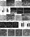
Micro-perforated basement membranes are present in multiple embryonic organs. (A) Brightfield images of an E13 SMG, E11 lung, and E11 kidney show the differences in morphology among developing branched organs; scale bars: 200 µm. (B) Maximum-intensity projection of an E13 SMG immunostained for collagen IV with labels indicating an epithelial bud with a developing cleft and its duct, surrounded by mesenchyme. Maximum projection images of confocal slices of E11 lung(C) and kidney (D) immunostained for laminin. Scale bars: 20 µm and 10 µm for the insets, respectively.
Fig. 2
Fig. 2
Characterization of basement membrane perforations. (A) Maximum projection images of an E13 SMG stained for several BM components: perlecan (magenta), laminin (green), collagen IV (cyan), plus merged; scale bar: 10 µm. (B) Maximum projection of E13.5 SMG stained for collagen IV marked with 4 rectangular regions of equal size for comparative analysis, starting at the tip of the bud and ending at the center of the bud; scale bar: 20 µm. (C) Average percent surface area of BM absent (± SEM) based on region: region 1 = tip of the bud and 4 = center of the bud, as shown in 2B. (D) Average number of holes per 500 µm2 area of BM, (E) average perforation areas, and (F) axial ratio of holes (± SEM) of each of the 4 different regions of the bud, ***p < 0.001.
Fig. 3
Fig. 3
Global and local basement membrane dynamics. (A) Rearward translocation of the BM as shown by the movement of a photobleached region rearward in 60 min. The dashed line indicates the starting point of the photobleached region. Scale bar: 10 µm. (B) Tracking of four (color-coded) perforations over 14 min in an E13 SMG; scale bar is 5 µm. (C) Average BM translocation velocity (± SEM) in glands with no treatment or treated with 50 µM blebbistatin or 5 µM BB-94 overnight, ***p < 0.001. (D) Change in area of two example holes in control and 50 µM blebbistatin-treated glands imaged over a span of 6 min. (E) Average distensibility of the BM (± SEM) expressed as maximum displacements within a 20-min assay period comparing a region near the tip versus mid-bud, or at the tip of glands treated with 50 µM blebbistatin or 5 µM BB-94, ***p < 0.001. (F) 50-s montage of a basal epithelial cell from an E13 GFP-myosin IIA gland (green) showing a cell process protruding (yellow arrowheads) through the BM (magenta) imaged with fluorescently tagged collagen IV antibody; scale bar: 5 µm.
Fig. 4
Fig. 4
Both protease and myosin II activity are required for basement membrane dynamics. (A) Control and (B) 5 µM BB-94 treated E12.5 glands immunostained with collagen IV after 24 h. Brightfield scale bar: 200 µm and collagen IV scale bar: 10 µm. (C) Fold change in BM intensity (± SEM) after 12 h of treatment with BB-94 compared with control in E13 SMGs. (D) Average bud outgrowth velocity (± SEM) after 12 h of treatment with BB-94, **p < 0.01 and ***p < 0.001. (E) GFP-myosin IIA and (F) IIB localization in an E13 SMG dissected from knock-in mice. Scale bar: 10 µm (G) Phospho-myosin light chain staining in an E12.5 epithelial rudiment cultured for 48 h in laminin gel. Scale bar: 10 µm (H) Control and (I) 50 µM blebbistatin-treated E12.5 glands after 24 h. Brightfield scale bar: 200 µm and collagen IV scale bar: 10 µm (J) E12.5 SMGs incubated overnight with or without 5 µM BB-94 or 50 µM blebbistatin and then incubated with EdU for 2 h. Scale bars: 100 µm. (K) Quantification of percent EdU positive nuclei in control and 5 µM BB-94 or 50 µM blebbistatin treated glands. (L) Quantification of percent DAPI per fixed area in control and 5 µM BB-94 or 50 µM blebbistatin treated glands. (M) E13 SMG stained for collagen IV after 2 h treatment with 50 µM blebbistatin, 5 µM BB-94, or both drugs together compared to control; scale bar: 5 µm.
Fig. 5
Fig. 5
Summary model. The basement membrane, displayed in magenta, surrounds the tips of expanding salivary gland epithelial end buds, and it becomes perforated with numerous holes that decrease in size and number closer to the middle of the bud. These micro-perforations are likely formed by a combination of proteases degrading the BM, epithelial cell protrusions extending through the BM (shown in green), as well as both local and global myosin II-mediated contraction of the BM. The presence of the micro-perforations increases the distensibility of the BM, allowing for outgrowth of the epithelium and myosin II-mediated rearward translocation and accumulation of the BM. Protease activity and actomyosin contractility are required for the local and global remodeling of the BM during development of the salivary gland.

References

    1. Andrew DJ, Ewald AJ. Morphogenesis of epithelial tubes: insights into tube formation, elongation, and elaboration. Dev. Biol. 2010;341:34–55. - PMC - PubMed
    1. Banerjee SD, Cohn RH, Bernfield MR. Basal lamina of embryonic salivary epithelia. Production by the epithelium and role in maintaining lobular morphology. J. Cell Biol. 1977;73:445–463. - PMC - PubMed
    1. Bernfield M, Banerjee SD. The turnover of basal lamina glycosaminoglycan correlates with epithelial morphogenesis. Dev. Biol. 1982;90:291–305. - PubMed
    1. Bluemink JG, Van Maurik P, Lawson KA. Intimate cell contacts at the epithelial/mesenchymal interface in embryonic mouse lung. J. Ultrastruct. Res. 1976;55:257–270. - PubMed
    1. Candiello J, Cole GJ, Halfter W. Age-dependent changes in the structure, composition and biophysical properties of a human basement membrane. Matrix Biol. 2010;29:402–410. - PubMed

Publication types

LinkOut - more resources