

Long-Term Consequences of Traumatic Brain Injury: Current Status of Potential Mechanisms of Injury and Neurological Outcomes
- PMID: 25158206
- PMCID: PMC4677116
- DOI: 10.1089/neu.2014.3352
Long-Term Consequences of Traumatic Brain Injury: Current Status of Potential Mechanisms of Injury and Neurological Outcomes
Abstract
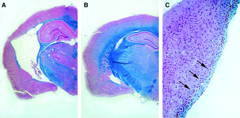
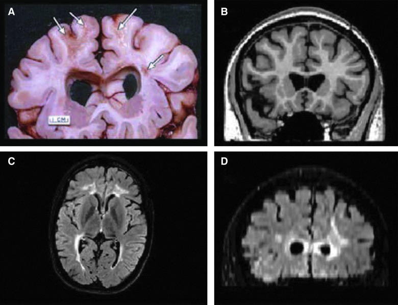
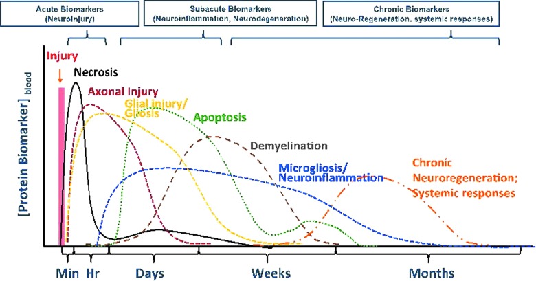
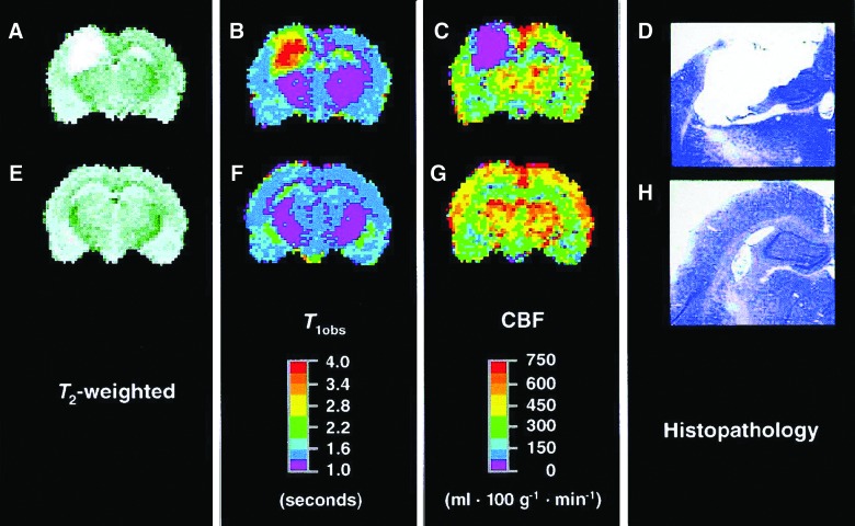
Traumatic brain injury (TBI) is a significant clinical problem with few therapeutic interventions successfully translated to the clinic. Increased importance on the progressive, long-term consequences of TBI have been emphasized, both in the experimental and clinical literature. Thus, there is a need for a better understanding of the chronic consequences of TBI, with the ultimate goal of developing novel therapeutic interventions to treat the devastating consequences of brain injury. In models of mild, moderate, and severe TBI, histopathological and behavioral studies have emphasized the progressive nature of the initial traumatic insult and the involvement of multiple pathophysiological mechanisms, including sustained injury cascades leading to prolonged motor and cognitive deficits. Recently, the increased incidence in age-dependent neurodegenerative diseases in this patient population has also been emphasized. Pathomechanisms felt to be active in the acute and long-term consequences of TBI include excitotoxicity, apoptosis, inflammatory events, seizures, demyelination, white matter pathology, as well as decreased neurogenesis. The current article will review many of these pathophysiological mechanisms that may be important targets for limiting the chronic consequences of TBI.
Keywords: TBI; atrophy; inflammation; neurogenesis; progressive damage; white matter.
Figures
References
-
- Masel B.E., and DeWitt D.S. (2010). Traumatic brain injury: a disease process, not an event. J. Neurotrauma 27, 1529–1540 - PubMed
-
- Bergeson A.G., Lundin R., Parkinson R.B., Tate D.F., Victoroff J., Hopkins R.O., and Bigler E.D. (2004). Clinical rating of cortical atrophy and cognitive correlates following traumatic brain injury. Clin. Neuropsychol. 18, 509–520 - PubMed
-
- Levin H.S., Amparo E., Eisenberg H.M., Williams D.H., High W.M., Jr., McArdle C.B., and Weiner R.L. (1987). Magnetic resonance imaging and computerized tomography in relation to the neurobehavioral sequelae of mild and moderate head injuries. J. Neurosurg. 66, 706–713 - PubMed
-
- Corrigan J.D., and Hammond F.M. (2013). Traumatic brain injury as a chronic health condition. Arch. Phys. Med. Rehabil. 94, 1199–1201 - PubMed
-
- Annegers J.F., Hauser W.A., Coan S.P., and Rocca W.A. (1998). A population-based study of seizures after traumatic brain injuries. N. Engl. J. Med. 338, 20–24 - PubMed
Publication types
MeSH terms
Grants and funding
LinkOut - more resources
Full Text Sources
Other Literature Sources
Medical
