. 2014 Nov:71:305-16.
doi: 10.1016/j.nbd.2014.08.022. Epub 2014 Aug 23.

Gabapentin attenuates hyperexcitability in the freeze-lesion model of developmental cortical malformation

Affiliations

Gabapentin attenuates hyperexcitability in the freeze-lesion model of developmental cortical malformation

Lauren Andresen et al. Neurobiol Dis. 2014 Nov.

Abstract

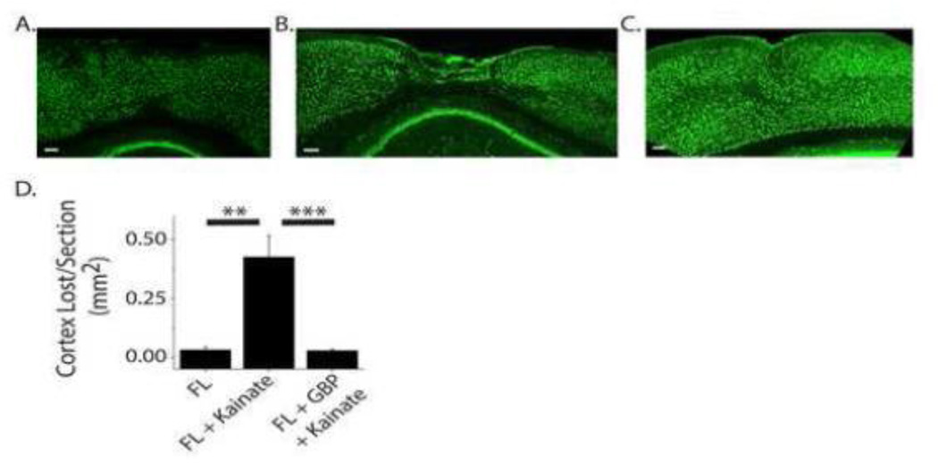
Developmental cortical malformations are associated with a high incidence of drug-resistant epilepsy. The underlying epileptogenic mechanisms, however, are poorly understood. In rodents, cortical malformations can be modeled using neonatal freeze-lesion (FL), which has been shown to cause in vitro cortical hyperexcitability. Here, we investigated the therapeutic potential of gabapentin, a clinically used anticonvulsant and analgesic, in preventing FL-induced in vitro and in vivo hyperexcitability. Gabapentin has been shown to disrupt the interaction of thrombospondin (TSP) with α2δ-1, an auxiliary calcium channel subunit. TSP/α2δ-1 signaling has been shown to drive the formation of excitatory synapses during cortical development and following injury. Gabapentin has been reported to have neuroprotective and anti-epileptogenic effects in other models associated with increased TSP expression and reactive astrocytosis. We found that both TSP and α2δ-1 were transiently upregulated following neonatal FL. We therefore designed a one-week GBP treatment paradigm to block TSP/α2δ-1 signaling during the period of their upregulation. GBP treatment prevented epileptiform activity following FL, as assessed by both glutamate biosensor imaging and field potential recording. GBP also attenuated FL-induced increases in mEPSC frequency at both P7 and 28. Additionally, GBP treated animals had decreased in vivo kainic acid (KA)-induced seizure activity. Taken together these results suggest gabapentin treatment immediately after FL can prevent the formation of a hyperexcitable network and may have therapeutic potential to minimize epileptogenic processes associated with developmental cortical malformations.

Keywords: Cortex; Developmental cortical malformation; Epilepsy; Freeze lesion; Gabapentin; Glutamate; Thrombospondin.


Figures

Figure 1
Figure 1. Thrombospondin and α2δ-1 immunoreactivity is increased after FL
(A) TSP1/2 staining in sham injured and FL cortex at P3, P7 and P14 (B) Average TSP1/2 fluorescence relative to sham (C) TSP1/2 (green) and GFAP (red) co-stain of a P7 FL animal and 60× magnification of the area in the black box for TSP1/2, GFAP and merged TSP1/2 (green) and GFAP (red) (D) α2δ-1 staining in sham injured and FL cortex at P3, P7 and P14 (E) Average α2δ-1 fluorescence relative to sham (F) α2δ-1 (red) and NeuN (green) co-stain of a P7 FL animal and 60× magnification of the area in the black box for α2δ-1, NeuN and merged α2δ-1 (red) and NeuN (green), *** P < 0.001, *P < 0.05.
Figure 2
Figure 2. Gabapentin treatment decreases network hyperexcitability following FL
(A) Representative evoked field excitatory post-synaptic potentials (fEPSPs) recorded in the neocortex from sham injured (black), FL (red) and GBP treated FL animals (gray). (B) Box-Whisker plot of percent of stimulus-evoked fEPSPs which had epileptiform activity in acute brain slices at threshold stimulation, ** P < 0.01, *** P < 0.001. (C) Box-Whisker plot of fEPSP area (integrated area under the curve 1s post-stimulation) at threshold stimulation, *P < 0.05 (D) Average fEPSP area at threshold and 2× threshold stimulation for sham, FL and GBP treated FL animals, *P < 0.05.
Figure 3
Figure 3. Gabapentin treatment decreases evoked extracellular glutamate signaling as measured using FRET based biosensor imaging
(A) Glutamate images of ΔFRET (CFP/Venus ratio) signal/ ΔFRET noise on a pixel-by-pixel basis at 100ms pre-stimulation, 400 ms post-stimulation and 1000 ms post-stimulation in sham, vehicle and GBP treated animals. Diagram illustrates cortical area being imaged with placement of recording electrode in cortical layer IV/V and stimulating electrode in the underlying white matter. (B) Individual ΔFRET signal/noise traces from sham (black), FL+Veh (red) and FL+GBP (gray) slices (C) and simultaneously recoreded evoked fEPSPs. (D) Average peak amplitude of ΔFRET signal/noise, ** P < 0.01.
Figure 4
Figure 4. An anticonvulsant with a different mechanism of action do not replicate the effects of gabapentin
(A) Example evoked cortical field potential from sham (black), FL (red) and phenobarbital treated FL (blue). (B) Epileptiform activity (%) in acute brain slices, ***P < 0.001 (C) fEPSP area, ** P < 0.01.
Figure 5
Figure 5. Gabapentin treatment decreases the frequency of excitatory postsynaptic currents in layer V pyramidal neurons of the FL cortex
(A) Representative mEPSC recording of layer V pyramidal neuron from Sham, FL and GBP treated FL cortex at P7. (B) Averaged mEPSC from sham (black), FL (gray) and GBP treated FL (blue) animals at P7. (C) Average mEPSC amplitude at P7. (D) Cumulative probability of inter-event intervals of FL and sham animals (P < 0.001 with D=0.6656) and FL and GBP treated FL animals at P7 (P < 0.001 with D=0.7848). (E) Representative mEPSC recording of layer V pyramidal neuron from Sham, FL and GBP treated FL cortex at P28. (F) Averaged mEPSC from sham (black), FL (gray) and GBP treated FL (blue) animals at P28. (G) Average mEPSC amplitude at P7, *P < 0.05. (H) Cumulative probability of inter-event intervals of FL and sham animals (P < 0.001 with D=0.3706) and FL and GBP treated FL animals at P28 (P < 0.001 with D=0.5994).
Figure 6
Figure 6. Gabapentin treatment decreases GFAP immunoreactivity following FL
(A) Representative images of GFAP staining of cortex from sham, vehicle treated FL and GBP treated FL animals at P7, P14 and P28. Scale bar = 100µm (B) Higher magnification of GFAP staining from microgyrus of P14 FL cortex (C) Percent of GFAP positive pixels in 200 µm ROI centered around lesion or equivalent cortical area, * P < 0.05, ** P < 0.01.
Figure 7
Figure 7. Gabapentin treatment decreases in vivo kainic acid-induced seizure activity
Representative power spectrum from 2 hour EEG recording of (A) sham injured, (B) FL and (C) GBP treated FL animals following acute kainic acid injection. (D) Percent time seizing and (E) average seizure duration, ** P < 0.01, *** P < 0.001. (F) Spectral analysis of EEG from sham (black), FL (red) and GBP treated FL animals (blue). Top, pre-kainic acid EEG, middle, kainic acid EEG, bottom, % baseline power normalized to pre-kainic acid EEG, * P < 0.05.
Figure 8
Figure 8. Gabapentin treatment protects against cortical tissue loss following acute kainic acid injection
Representative images of NeuN staining of cortex of (A) FL animal treated with vehicle, (B) FL animal treated with kainic acid and (C) GBP treated FL animal treated with kainic acid. Scale bar = 100 µm. (D) Average cortical tissue lost per section, ** P < 0.01, *** P < 0.001.

References

    1. Adams JC, Lawler J. The thrombospondins. Int J Biochem Cell Biol. 2004;36:961–968. - PMC - PubMed
    1. Asch AS, Leung LL, Shapiro J, Nachman RL. Human brain glial cells synthesize thrombospondin. Proc Natl Acad Sci U S A. 1986;83:2904–2908. - PMC - PubMed
    1. Bordey A, Lyons SA, Hablitz JJ, Sontheimer H. Electrophysiological characteristics of reactive astrocytes in experimental cortical dysplasia. J Neurophys. 2001;85:1719–1731. - PubMed
    1. Brill J, Huguenard JR. Enhanced infragranular and supragranular synaptic input onto layer 5 pyramidal neurons in a rat model of cortical dysplasia. Cereb Cortex. 2010;20:2926–2938. - PMC - PubMed
    1. Campbell SL, Hablitz JJ. Decreased glutamate transport enhances excitability in a rat model of cortical dysplasia. Neurobiol Dis. 2008 - PMC - PubMed

Publication types

MeSH terms