. 2014 Aug 27:11:139.
doi: 10.1186/s12974-014-0139-x.

Longitudinal follow-up of autophagy and inflammation in brain of APPswePS1dE9 transgenic mice

Affiliations

Longitudinal follow-up of autophagy and inflammation in brain of APPswePS1dE9 transgenic mice

Arnaud François et al. J Neuroinflammation. .

Abstract

Background: In recent years, studies have sought to understand the mechanisms involved in the alteration of autophagic flux in Alzheimer's disease (AD). Alongside the recent description of the impairment of lysosomal acidification, we wanted to study the relationships between inflammation and autophagy, two physiological components deregulated in AD. Therefore, a longitudinal study was performed in APPswePS1dE9 transgenic mice at three, six and twelve months of age.

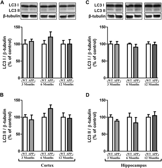
Methods: Autophagic markers (Beclin-1, p62 and LC3) and the activation of mammalian Target of Rapamycin (mTOR) signaling pathway were quantified by western blot. Cytokine levels (IL-1β, TNF-α and IL-6) were measured by ELISA. Transmission electron microscopy was performed to detect autophagic vacuoles. Mann-Whitney tests were used to compare wild-type (WT) versus APPswePS1dE9 mice. Longitudinal changes in parameters were analyzed with a Kruskal-Wallis test followed by a post-hoc Dunn's test. Correlation between two parameters was assessed using a Spearman test.

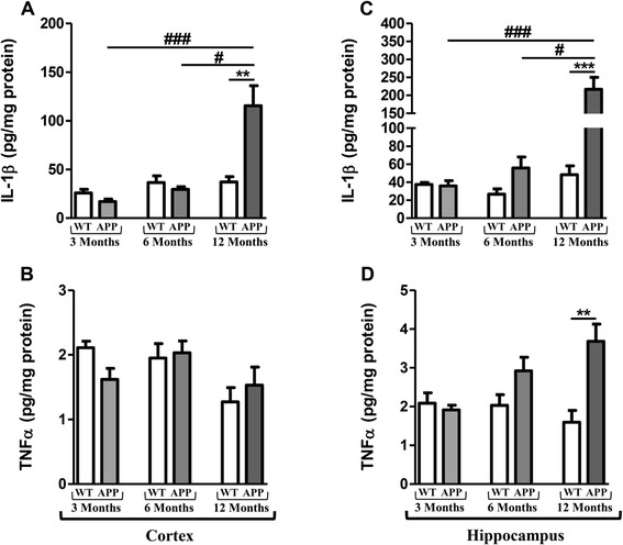
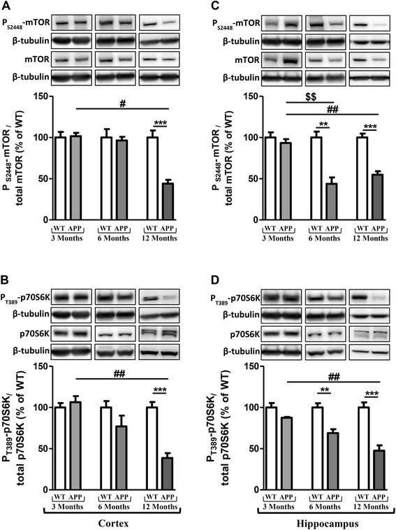
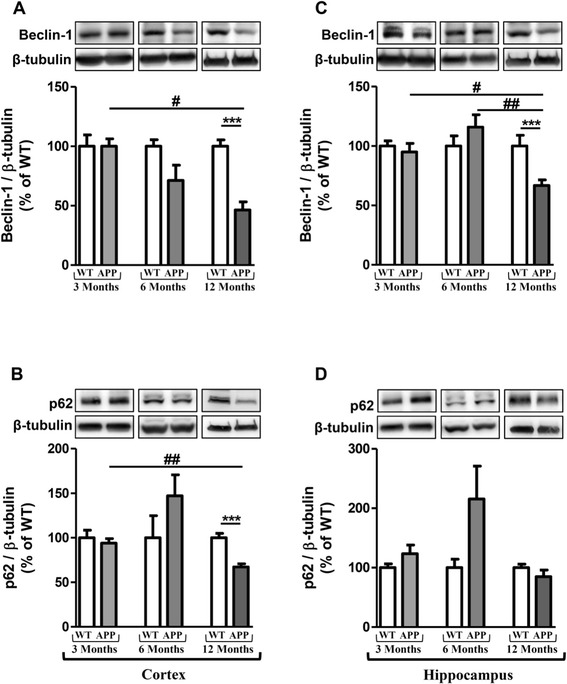
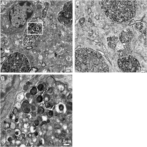
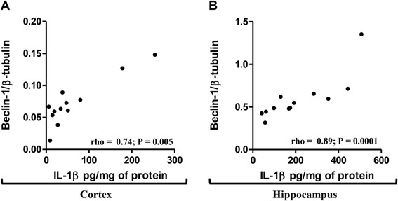
Results: Compared to 12-month old WT mice, 12-month old APPswePS1dE9 mice had higher levels of IL-1β and TNF-α, a greater inhibition of the mTOR signaling pathway and lower levels of Beclin-1 expression both in cortex and hippocampus. Regarding the relationship of the various parameters in 12-month old APPswePS1dE9 mice, Beclin-1 rates were positively correlated with IL-1β and TNF-α levels. And, on the contrary, TNF-α levels were inversely correlated with the levels of mTOR activation. Altogether, these results suggest that inflammation could induce autophagy in APPswePS1dE9 mice. However, these transgenic mice displayed a large accumulation of autophagic vesicles within dystrophic neurons in cortex and hippocampus, indicating a terminal failure in the autophagic process.

Conclusions: This first demonstration of relationships between inflammation and autophagy in in vivo models of AD should be taken into account in new therapeutic strategies to prevent inflammation and/or stimulate autophagy in advanced neurodegenerative process such as AD.


Figures

Figure 1
Figure 1
Longitudinal monitoring of cytokines in the brain of APPswePS1dE9 mice. IL1-β and TNF-α levels were measured by ELISA in cortex (A and B) and hippocampus (C and D) of APPswePS1dE9 (APP) and wild-type mice (WT) at three, six and twelve months of age. Cytokine levels were expressed in pg/mg protein. Results are mean ± SEM of 12 mice in each group. Comparison between APP and WT mice at each age by a Mann-Whitney test: ** P < 0.01, *** P < 0.001; comparison of the 3 classes of age (Kruskall-Wallis test) followed by two-by-two comparisons (Dunn’s multiple comparison test in APP mice): # P < 0.05 for 6-month old versus 12-month old APP mice, ### P < 0.001 for 3-month old versus 12-month old APP mice.
Figure 2
Figure 2
Changes in activation of the mammalian Target of Rapamycin (mTOR) signaling pathway. Representative immunoblots of PS2448-mTOR (289 kDa), total mTOR (289 kDa), PT389-p70S6K (70 kDa), total p70S6K (70 kDa) in cortex (A and B) and hippocampus (C and D) of wild-type (WT) mice and APPswePS1dE9 (APP) at 3, 6 and 12 months of age. Semi-quantitative analysis of immunoblot was performed using Gene Tools software (Syngene, Ozyme, Saint-Quentin-en-Yvelines, France). The immunoreactivity of protein was normalized to β-tubulin (55 kDa) immunoreactivity. The results are expressed as arbitrary units (% of WT). Results are mean ± SEM of 12 mice. Comparison between APP and WT mice at each age by a Mann-Whitney test: ** P < 0.01, *** P < 0.001; comparison of the 3 classes of age (Kruskall-Wallis test) followed by two-by-two comparisons (Dunn’s multiple comparison test in APP mice): $$ P < 0.01 for 3-month old versus 6-month old APP mice, # P < 0.05, ## P < 0.01 for 6-month old versus 12-month old APP mice.
Figure 3
Figure 3
Longitudinal monitoring of Beclin-1 and p62 in the brain of APPswePS1dE9 mice. Representative immunoblots of Beclin-1 (60 kDa) and p62 (62 kDa) in cortex (A and B, respectively) and hippocampus (C and D, respectively) of wild-type (WT) and APPswePS1dE9 (APP) mice at 3, 6 and 12 months of age. Semi-quantitative analysis of immunoblot was performed using Gene Tools software (Syngene, Ozyme, Saint-Quentin-en-Yvelines, France). The immunoreactivity of protein was normalized to β-tubulin (55 kDa) immunoreactivity. The results are expressed as arbitrary units (% of WT). Results are mean ± SEM of 12 mice. Comparison between APP and WT mice at each age by a Mann-Whitney test: *** P < 0.001; Comparison of the three classes of age (Kruskall-Wallis test) followed by two-by-two comparisons (Dunn’s multiple comparison test in APP mice): # P < 0.05, ## P < 0.01 compared to 12-month old APPswePS1dE9.
Figure 4
Figure 4
Longitudinal monitoring of LC3 in the brain of APPswePS1dE9 mice. Representative immunoblots of LC3-I (18 kDa), LC3-II (16 kDa) in cortex (A and B, respectively) and hippocampus (C and D, respectively) of wild-type (WT) and APPswePS1dE9 (APP) mice at 3, 6 and 12 months of age. Semi-quantitative analysis of immunoblot was performed using Gene Tools software (Syngene, Ozyme, Saint-Quentin-en-Yvelines, France). The immunoreactivity of protein was normalized to β-tubulin (55 kDa) immunoreactivity. The results are expressed as arbitrary units (% of WT). Results are mean ± SEM for 12 mice.
Figure 5
Figure 5
Ultrastructure of cortex and hippocampus in 12-month old APPswePS1dE9 mice. Transmission electron microscopy (TEM) cortical (A) and hippocampal (C) representative images (n = 3 mice) showed dystrophic neurites (marked by *) with a large accumulation of autophagic vesicles (AVs). (B) represents a magnified region of interest (defined by a white square in image (a)) within many AVs with a dense compacted amorphous or multilamellar content as indicated by white arrows.
Figure 6
Figure 6
Correlation between Beclin-1 and IL-1β levels in cortex and hippocampus of 12-month old APPswePS1dE9 mice. Spearman correlations were performed between levels of IL-1β and Beclin-1 in the cortex (A) and hippocampus (B) of 12-month old APPswePS1dE9 mice (n = 12). rho and P-values were indicated on graphs. The level of significance was P < 0.05.

References

    1. Abraham CR, Selkoe DJ, Potter H. Immunochemical identification of the serine protease inhibitor alpha 1-antichymotrypsin in the brain amyloid deposits of Alzheimer's disease. Cell. 1988;52:487–501. doi: 10.1016/0092-8674(88)90462-X. - DOI - PubMed
    1. Luber-Narod J, Rogers J. Immune system associated antigens expressed by cells of the human central nervous system. Neurosci Lett. 1988;94:17–22. doi: 10.1016/0304-3940(88)90263-7. - DOI - PubMed
    1. McGeer PL, Itagaki S, McGeer EG. Expression of the histocompatibility glycoprotein HLA-DR in neurological disease. Acta Neuropathol. 1988;76:550–557. doi: 10.1007/BF00689592. - DOI - PubMed
    1. Rogers J, Luber-Narod J, Styren SD, Civin WH. Expression of immune system-associated antigens by cells of the human central nervous system: relationship to the pathology of Alzheimer's disease. Neurobiol Aging. 1988;9:339–349. doi: 10.1016/S0197-4580(88)80079-4. - DOI - PubMed
    1. Rodriguez JJ, Olabarria M, Chvatal A, Verkhratsky A. Astroglia in dementia and Alzheimer's disease. Cell Death Differ. 2009;16:378–385. doi: 10.1038/cdd.2008.172. - DOI - PubMed

Publication types

MeSH terms

LinkOut - more resources