. 2014;9(4):437-52.
doi: 10.2217/rme.14.26.

Immunohistochemical toolkit for tracking and quantifying xenotransplanted human stem cells

Affiliations

Immunohistochemical toolkit for tracking and quantifying xenotransplanted human stem cells

Justine Allard et al. Regen Med. 2014.

Abstract

Aim: Biomarker-based tracking of human stem cells xenotransplanted into animal models is crucial for studying their fate in the field of cell therapy or tumor xenografting.

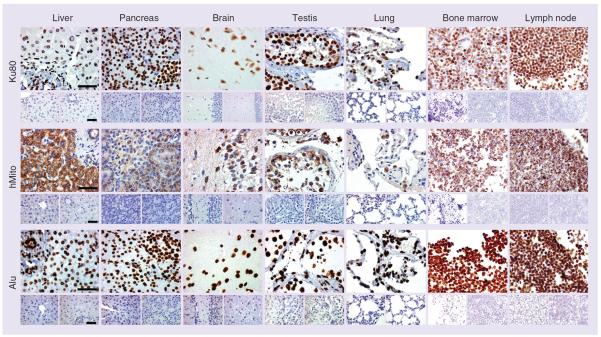
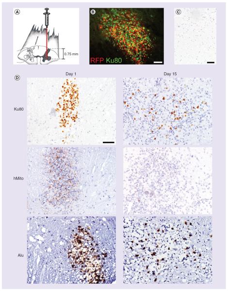
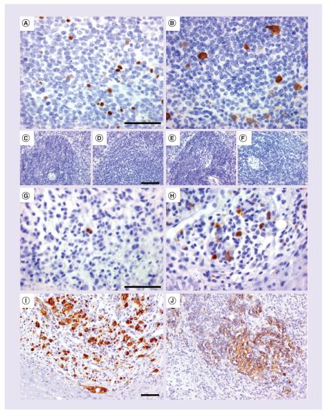
Materials & methods: Using immunohistochemistry and in situ hybridization, we analyzed the expression of three human-specific biomarkers: Ku80, human mitochondria (hMito) and Alu.

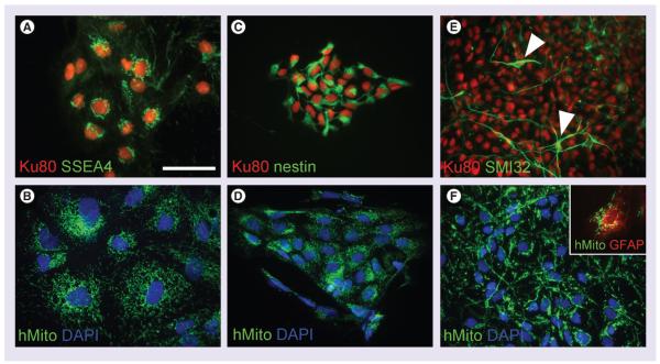
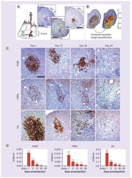
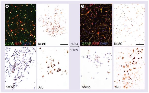
Results: We showed that Ku80, hMito and Alu biomarkers are broadly expressed in human tissues with no or low cross-reactivity toward rat, mouse or pig tissues. In vitro, we demonstrated that their expression is stable over time and does not change along the differentiation of human-derived induced pluripotent stem cells or human glial-restricted precursors. We tracked in vivo these cell populations after transplantation in rodent spinal cords using aforementioned biomarkers and human-specific antibodies detecting apoptotic, proliferating or neural-committed cells.

Conclusion: This study assesses the human-species specificity of Ku80, hMito and Alu, and proposes useful biomarkers for characterizing human stem cells in xenotransplantation paradigms.

Keywords: cell therapy; human-specific biomarker; image quantification; stem cells; xenotransplantation.


Figures

Figure 1
Figure 1. Immunohistochemistry of Ku80, human mitochondria and Alu biomarkers in organs from human, mouse and rat origins
All three biomarkers are expressed in human liver, pancreas, brain, testis, lung, hematopoietic bone marrow and lymph node (upper panels) but not in mouse (lower left panels) or rat (lower right panels) tissue counterpart. Ku80 and Alu strongly label the nucleus while hMito is confined to the cytoplasmic compartment. In most of the human organs, more than 99% of the cells, regardless of their tissular origin (epithelial, muscular, connective), are immunoreactive for Alu and Ku80 biomarkers. hMito is less broadly expressed and for instance does not label all types of human pneumocytes. A detailed analysis of biomarker expression is shown in Table 2. Scale bar represents 50 μm. hMito: Human mitochondria.
Figure 2
Figure 2. Immunohistochemistry of Ku80 and human mitochondria along the differentiation process of human induced pluripotent stem cells toward neuronal lineage
(A & B) Ku80 and hMito immunolabeled all clones of induced pluripotent stem cell, whose cells are characterized by the expression of the SSEA4. (C & D) Upon differentiation into neural precursor cells, they start expressing nestin while keeping Ku80 and hMito expression. (E & F) At further differentiation stages, cells with a typical neuronal morphology were observed (white arrowheads in E). SMI32+ neurons were still expressing Ku80 and hMito. Sparse GFAP+ cells were also found immunoreactive for hMito (inset in F). Scale bar represents 50 μm. DAPI: 4′,6-diamidino-2-phenylindole; hMito: Human mitochondria.
Figure 3
Figure 3. Human neural precursor cells are injected into the rat spinal cord, targeting the ventral horn (facing page)
(A) At 1-day postinjection, Ku80, hMito and Alu immunostainings help visualize the injection site. (B) Computer-assisted image analysis allows to make the difference between the blue staining from hematoxylin counterstaining and the di-amino-benzidine brown precipitates from specific anti-human immunohistochemistry. Hence, LI corresponding to the percentage of the immunostained tissue surface was calculated. (C) Using anti-human Ku80, hMito and Alu immunohistochemistry, neural precursor cell-derived human cells are visualized in the spinal tissue from 1 day to 60 days post-transplantation. (D) Around the injection site, we detect inflammatory reaction and gliosis, most likely causing a loss of engrafted human cells over time. This is confirmed by image quantification showing a progressive decrease of the LI of Ku80, Alu and hMito immunostainings. Scale bars represent 500 μm in (A) and 100 μm in (C). Results are expressed as mean ± standard error of the mean. *p < 0.05, **p < 0.01, ***p < 0.001 for comparisons 1, 5, 15, 30 and 60 days versus sham group. The number of animals was, respectively, 8, 3, 5, 7, 14 and 5 rats for the experimental conditions: sham, 1, 5, 15, 30 and 60 days. hMito: Human mitochondria; LI: Labeling index.
Figure 4
Figure 4. Immunohistochemistry of Ku80, human mitochondria and Alu along the differentiation process of human glial-restricted precursors toward astrocytic lineage
(A) Undifferentiated glial-restricted precursors (GRP) are isolated from human fetuses based on their A2B5-specific expression. Cells are further transduced with a retroviral vector driving the RFP reporter gene. Undifferentiated GRP preferentially display a rounded shape and express all three biomarkers: Ku80, hMito and Alu. (B) Upon differentiation with BMP4, star-shaped cells are observed close to an astrocyte morphology and showing a strong immunoreactivity for GFAP. Differentiated GRP still express Ku80 and Alu, but the immunostaining for hMito is more faint. Scale bar represents 50 μm. hMito: Human mitochondria; RFP: Red fluorescent protein.
Figure 5
Figure 5. Detection of human glial-restricted precursors injected into the mouse spinal cord
(A) RFP prelabeled human glial-restricted precursors (GRP) are injected into the mouse spinal cord, targeting the ventral horn. (B) At 1-day postinjection, RFP+ human GRPs are visualized in the injection site of the spinal cord. These cells co-express the human-specific biomarker Ku80. (C) Using anti-human Ku80, hMito and Alu immunohistochemistry, human GRP cells can be tracked in the spinal tissue from 1 day to 15 days post-transplantation. (D) Vehicle-injected animals do not show any biomarker immunoreactivity in the spinal tissue. While Ku80 and Alu immunoreactive cells are still found at 15 days post-transplantation, the signal for hMito is very weak. Scale bar represents 50 μm. hMito: Human mitochondria; RFP: Red fluorescent protein.
Figure 6
Figure 6. Human biomarkers of apoptosis, proliferation and neural commitment
(A) c-PARP and (B) CyclinB1 antibodies are biomarkers detecting cell death and proliferation in human spleen, which do not cross-react with (C–E) mouse or (D–F) rat spleens. (G) Fifteen days post-transplantation of human neural precursor cells, these detected cells undergoing apoptosis or (H) clusters of proliferating human cells into the rat spinal cord tissue. (I) Neural precursor cells express nestin at 1-day postinjection and (J) later on become progressively immunoreactive for a human-specific neuron-specific enolase antibody. Scale bar represents 50 μm.

References

    1. Baddour JA, Sousounis K, Tsonis PA. Organ repair and regeneration: an overview. Birth Defects Res. C Embryo Today. 2012;96(1):1–29. - PubMed
    1. Lindvall O, Kokaia Z. Stem cells in human neurodegenerative disorders – time for clinical translation? J. Clin. Invest. 2010;120(1):29–40. - PMC - PubMed
    1. Mitrecic D, Nicaise C, Klimaschewski L, Gajovic S, Bohl D, Pochet R. Genetically modified stem cells for the treatment of neurological diseases. Front. Biosci. (Elite Ed.) 2012;4:1170–1181. - PubMed
    1. Glover JC, Boulland JL, Halasi G, Kasumacic N. Chimeric animal models in human stem cell biology. ILAR J. 2009;51(1):62–73. - PubMed
    1. Bengel FM. Noninvasive stem cell tracking. J. Nucl. Cardiol. 2011;18(5):966–973. - PubMed

Publication types

Substances